In the first of a two-part series, Derek Hampton introduces Shrivastava and colleagues’ (2021) review, delving into dental biofilm management and begins to explore a new protocol – guided biofilm therapy.
Dental biofilm is a polymicrobial entity that resides on biotic and abiotic surfaces of the oral cavity (Sanz et al, 2017). These include hard and soft tissues of the oral cavity. Also, surfaces such as orthodontic bands, clear aligners, or prostheses (Meto et al, 2019; Lasserre et al, 2018).
Supra and subgingival dental plaque biofilms can form on the tooth or implant surface. Being close to the gingival epithelium, can negatively affect periodontal and peri-implant health (Lasserre et al, 2018).
Dental plaque biofilms also form in some regions of the oral cavity from which it is difficult to remove, thus compromising home-care oral hygiene management.
Scaling and root planing (SRP) is considered the gold standard for mechanical plaque debridement (Lindhe et al, 1984). However, it also has disadvantages (Sultan et al, 2017; Rabbani et al, 1981; Eaton et al, 1985).
Nowadays, an alternative novel approach is being practised for removal of the biofilm by visualising it with a disclosing agent and subsequently removing it with specialised air abrasive powder. It is then followed by the removal of supra and subgingival calculus using specialised instruments. This concept has been named guided biofilm therapy (Mensi et al, 2020).
Dental biofilm and gum diseases
The oral cavity is inhabited by many microbial species, ranging from healthy microorganisms to those with pathogenic potential.
The association between dental plaque and periodontal diseases is a well-established fact.
However, until 1980, it was believed that the microorganism present in dental plaque remains in suspended or planktonic states (Seneviratne et al, 2011).
In line with this, the majority of treatment was directed towards the removal of dental plaque. Some time later, research indicated that the microorganisms are not free-floating entities; rather, they are attached to the tooth surfaces (Seneviratne et al, 2011).
It is now widely accepted that the microorganisms live in a complex environment known as biofilm (Seneviratne et al, 2011; Marsh, Zaura, 2017). It is known to be an aetiological factor for dental caries and periodontal disease development (Marsh and Zaura, 2017; Nimbulkar et al, 2020).
A mature biofilm is a polymicrobial entity that primarily consists of bacteria. Although, it can also harbour protozoa, viruses, and fungi (Larsen and Fiehn, 2017).
In 2002, Donlan and Casterton defined biofilm as a sessile microbiological community characterised by cells adhered to a substrate, to an interface, or to each other, embedded in an extracellular polymeric substance matrix that produces and presents an altered phenotype, in terms of growth rate and gene transcription (Rode et al, 2012).
Biofilm microorganisms display characteristics as a whole unit rather than as individual entities (Berger et al, 2014).
Dysbiosis
Usually, the bacteria residing in the biofilm are considered beneficial. However, during diminished host response predisposed by certain clinical situations, there is a shift in the composition of microbial flora. Pathogenic bacterial species dominate over the healthy microbial flora.
This phenomenon is known as ‘dysbiosis’ (Lasserre et al, 2018).
The bacteria residing in the biofilm are responsible for the inflammatory cascade and, subsequently, the destruction of the supporting tissues (Shrivastava et al, 2021; Shrivastava et al, 2020].
Currently, periodontal and peri-implant diseases are considered to be based on ‘polymicrobial synergy and dysbiosis’ (Hajishengallis and Lamont, 2012).
This has been established based upon the hypothesis that keystone pathogens such as P. gingivalis are initially introduced into the biofilm.
Later, by undermining the host’s immunity, they succeed in modifying the composition of the microbial community. This makes it more pathogenic and able to trigger disease (Hajishengallis et al, 2012).
Microbiota
These microbial changes are heightened by local environmental changes, creating microbiota capable of sustaining dysbiosis and progressing disease.
It is also suggested that instead of directly causing the disease, the keystone pathogens bring about a change in the metabolic activity of the commensal traits, which, in turn, increases the pathogenicity of the bacteria and manifests in the form of periodontal or peri-implant disease (Darveau, 2010).
The dysbiosis leads to an upsurge in the generation of inflammatory mediators, which triggers the host cell to produce toxic products.
When these toxic products exceed the threshold level, it leads to destruction of the tissues around the tooth or implant (Lasserre et al, 2018).
Additionally, the pathogenic bacteria trigger the innate immune response, which attempts to purify the invading microorganism (Silva et al, 2015).
In the innate immune system, the pathogens trigger the pattern recognition receptors (PRRs) that attach to the pathogen-associated molecular patterns (PAMPs). These receptor types include toll-like receptors, nucleotide-binding oligomerisation domain (NOD) proteins, cluster of differentiation 14 (CD14), complement receptor-3, lectins, and scavenger receptors (Lasserre et al, 2018; Amano, 2010).
Protein pathways
The toll-like receptors play a crucial role in the progression of periodontal/peri-implant inflammation and bone resorption (Kajiva and Kurihara, 2021).
It has been reported that PAMPs activates T and B cells’ immune response, leading to activation of cytokines and an osteolytic pathway (Kajiva and Kurihara, 2021).
In conjunction with innate immunity, periodontal and peri-implant tissue produces various cytokines and chemokines, which maintain the equilibrium. However, in the presence of dysbiosis, there are certain cytokines – such as IL-1β, tumour necrosis factor (TNF)-α and IL-6 – that lead to destruction of the tissues (Lasserre et al, 2018).
Apart from these mechanisms, there are three protein pathways, namely nuclear factor kappa B (NF-кB), cyclo-oxygenase (COX) and lipo-oxygenase (LOX), which has an established role in the progression of periodontal and/or peri-implant diseases (Lasserre et al, 2018).
Therefore, understanding its structure and biology is fundamental to discovering the cause and development of periodontal and peri-implant diseases.
For example, biofilm formed on the natural tooth or dental implant shares a common pattern of microbial colonisation (Dihr, 2013).
Biofilm formation is an inevitable phenomenon. However, its control and elimination must not be overlooked, as it is one of the main causes of periodontal and peri-implant diseases.
The need for effective hygiene measures
Dental biofilm resides in close vicinity to the oral gingival epithelium. Should oral hygiene measures prove ineffective, this supragingival biofilm will accumulate along the gingival epithelium. It could become a potential source of gingival inflammation (Lasserre, 2018; Shrivastava et al, 2021).
It is generally considered that the dental biofilm is harmful in nature. If not disrupted, it may progress to periodontitis, provided there is a simultaneous diminished host response (Sahni et al, 2016; Fatima et al, 2021).
To maintain periodontal stability following non-surgical or surgical periodontal, supportive periodontal therapy (SPT) plays an important role (Ng, 2018).
It is commonly observed that periodontal pockets can be easily recolonised with bacteria. This means regular recall visits in the form of periodontal maintenance therapy are of the utmost importance (Renvert and Persson, 2004).
In addition, the biofilm formed on the dental implant has a similar microbiota as the adjacent tooth (Cortés-Acha et al, 2017).
Periodontal disease
It has been observed that the subgingival microbiota share common periodontal pathogens as in periodontal disease. Therefore, maintenance of the implant by removing the biofilm should be the principal management to combat the development of peri-mucositis or peri-implantitis.
Oral hygiene is maintained at home via personal care. This includes the use of a toothbrush with toothpaste (Sahni et al, 2016; Meto et al, 2020). However, despite meticulous cleaning, some amount of dental biofilm may be left behind in undetected areas.
Dental anatomical structures – such as furcation, cervical enamel projection, deep grooves and concavities – can provide a potential biological recess for bacteria (Park et al, 2018).
Professional management of dental biofilm will enable professionals to reach inaccessible areas where dental plaque remains hidden.
SRP is a gold standard in non-surgical mechanical debridement, based on the biofilm’s mechanical disruption (Lasserre et al, 2018).
Although it is a conventional treatment option, it has its own disadvantages such as being a time-consuming procedure, technically demanding and occasionally uncomfortable for patients (Fleischer et al, 2015).
SRP
In addition, following SRP, it has been reported that the lingual tooth surface and furcation areas are prone to residual calculus (Rabbani et al, 1981; Eaton et al, 1985).
Moreover, furcation areas have been shown to have incomplete root planing (Eaton et al, 1985; Fischer et al, 1991).
Gingival recession and irreversible root damage have also been reported if SRP is performed repeatedly. This is a protocol for supportive periodontal therapy (Sultan et al, 2017).
These troublesome consequences may lead to dentinal hypersensitivity (Greenstein, 1992).
Furthermore, it has been observed that the outcome of SRP also depends on the skill level of the clinician (Boyd et al, 2016).
Considering these drawbacks, various technologies have been introduced to remove the dental biofilm, such as air polishing.
Introducing guided biofilm therapy
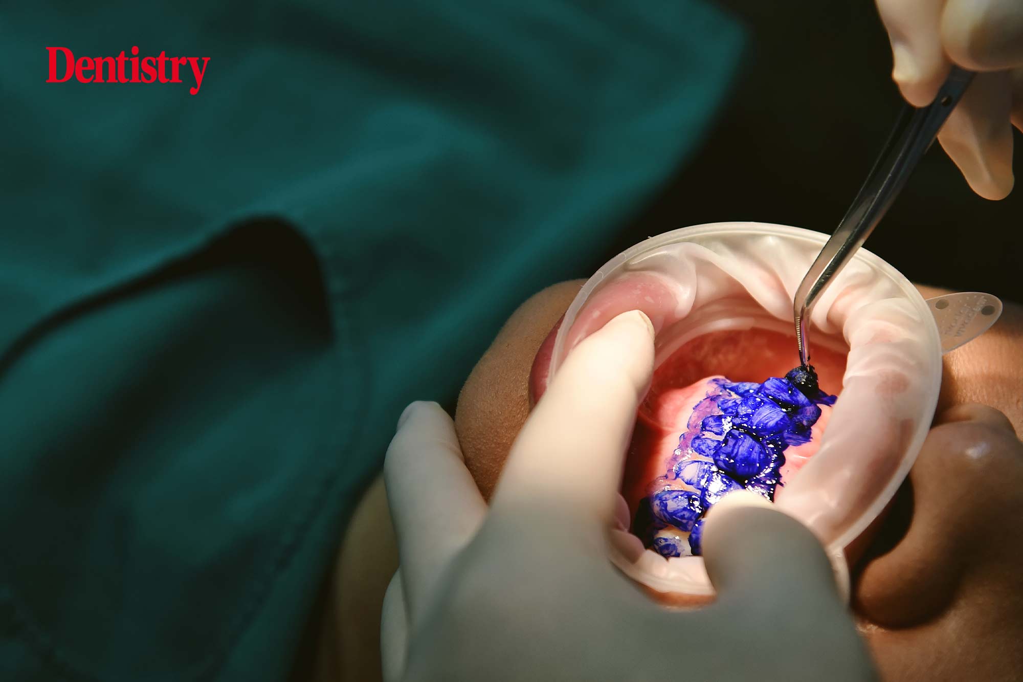
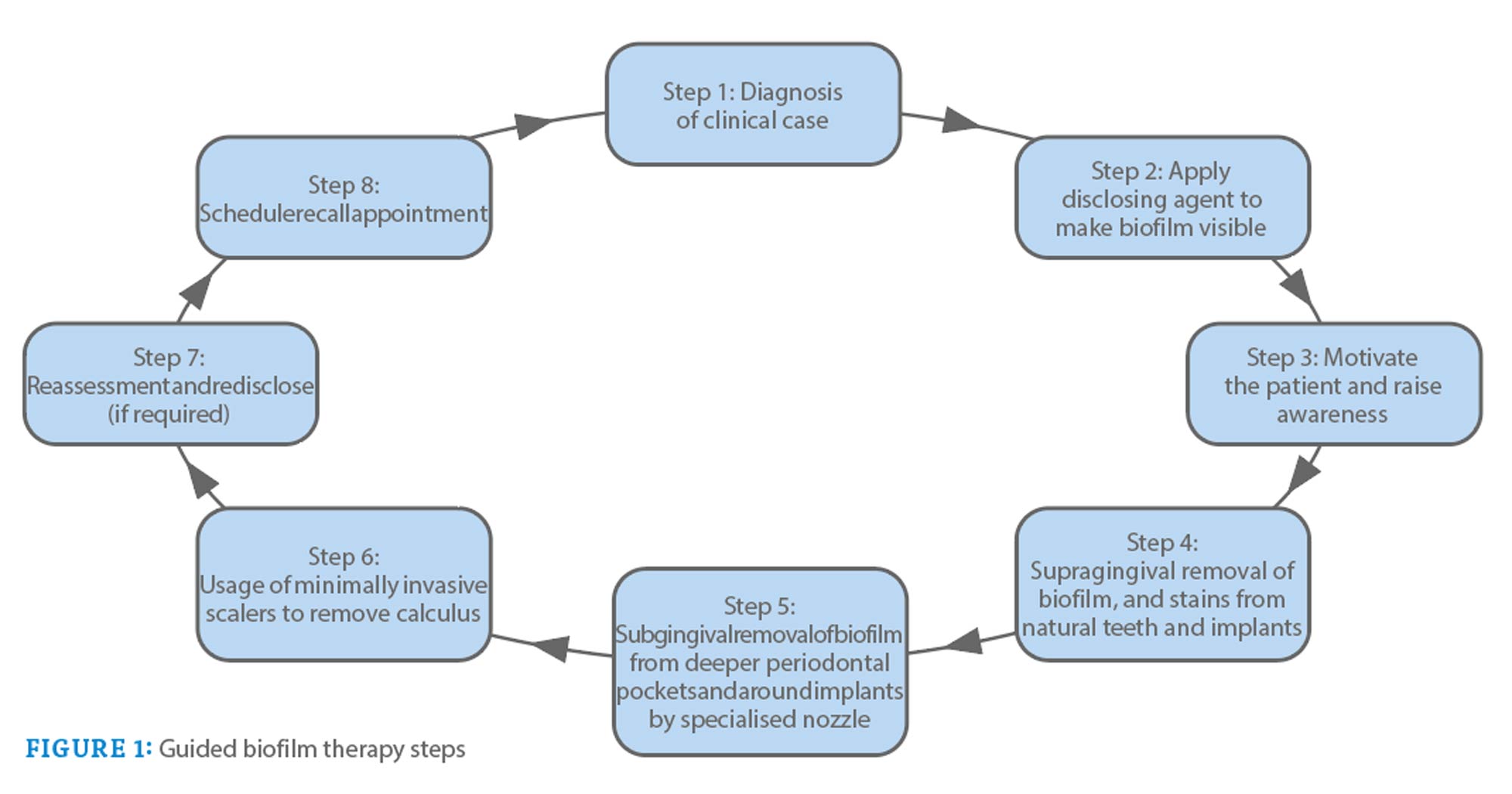
Guided biofilm therapy (GBT) is a new regimen that follows a sequential protocol for the removal of plaque and calculus. It starts with the detection of biofilm using a disclosing agent. It is followed by the use of air abrasive powder for the removal of plaque and stains.
Subgingival plaque and calculus are then removed with a specialised nozzle an. If required, eventual scaling with a specialised tip is performed.
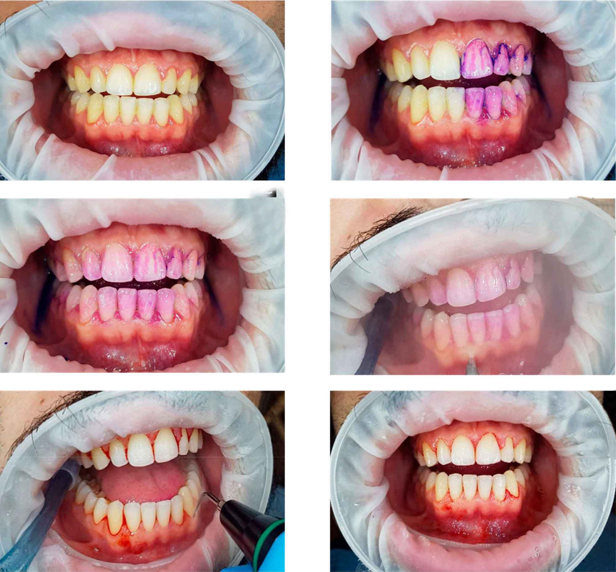
The sequential steps of GBT are described in Figure 1. Figure 2 illustrates the procedure performed on a patient with generalised bleeding on probing, plaque accumulation and localised calculus.
This review will continue in the next issue. There will be a particular focus on the GBT process and the supporting evidence base.
This article appeared in Clinical Dentistry. You can sign up to the magazine here.

