Here, we are taken through the treatment of an unerupted supernumerary tooth in a mesioangular position.
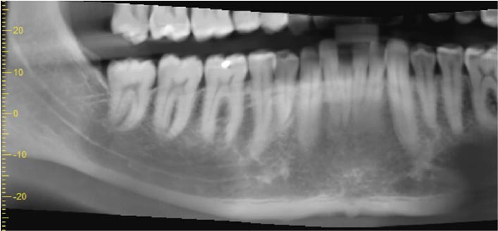
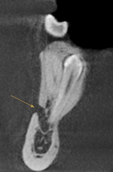
There is an unerupted supernumerary tooth lying in a mesioangular position, and with a lingual inclination, in the right premolar region of the mandible (Figure 1). It is 19.5mm in length.
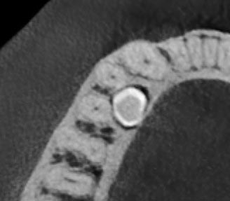
The crown of the supernumerary tooth is lingual to the middle thirds of the roots of LR4 (44) and LR5 (45), below a thinned lingual cortical plate (Figure 3). The pericoronal follicle space is of normal size.
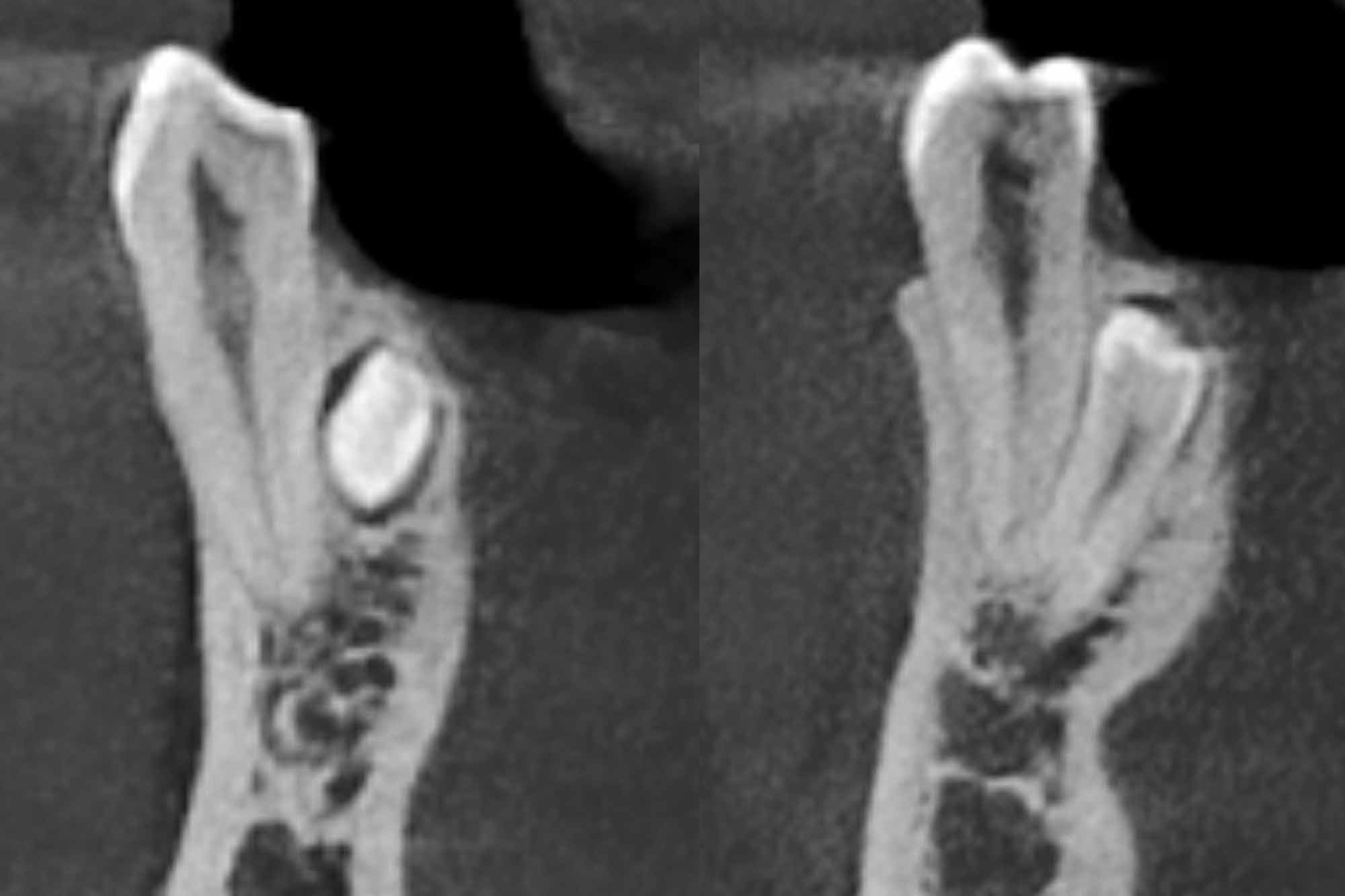
The crown of the supernumerary tooth is impacted against the middle third of the root of LR4 (44) (Figure 4a), while both the crown and root are in contact with the middle and apical thirds of the root of LR5 (45) (Figure 4b).
Surgical context
There is no root resorption seen on LR4 (44) or LR5 (45).
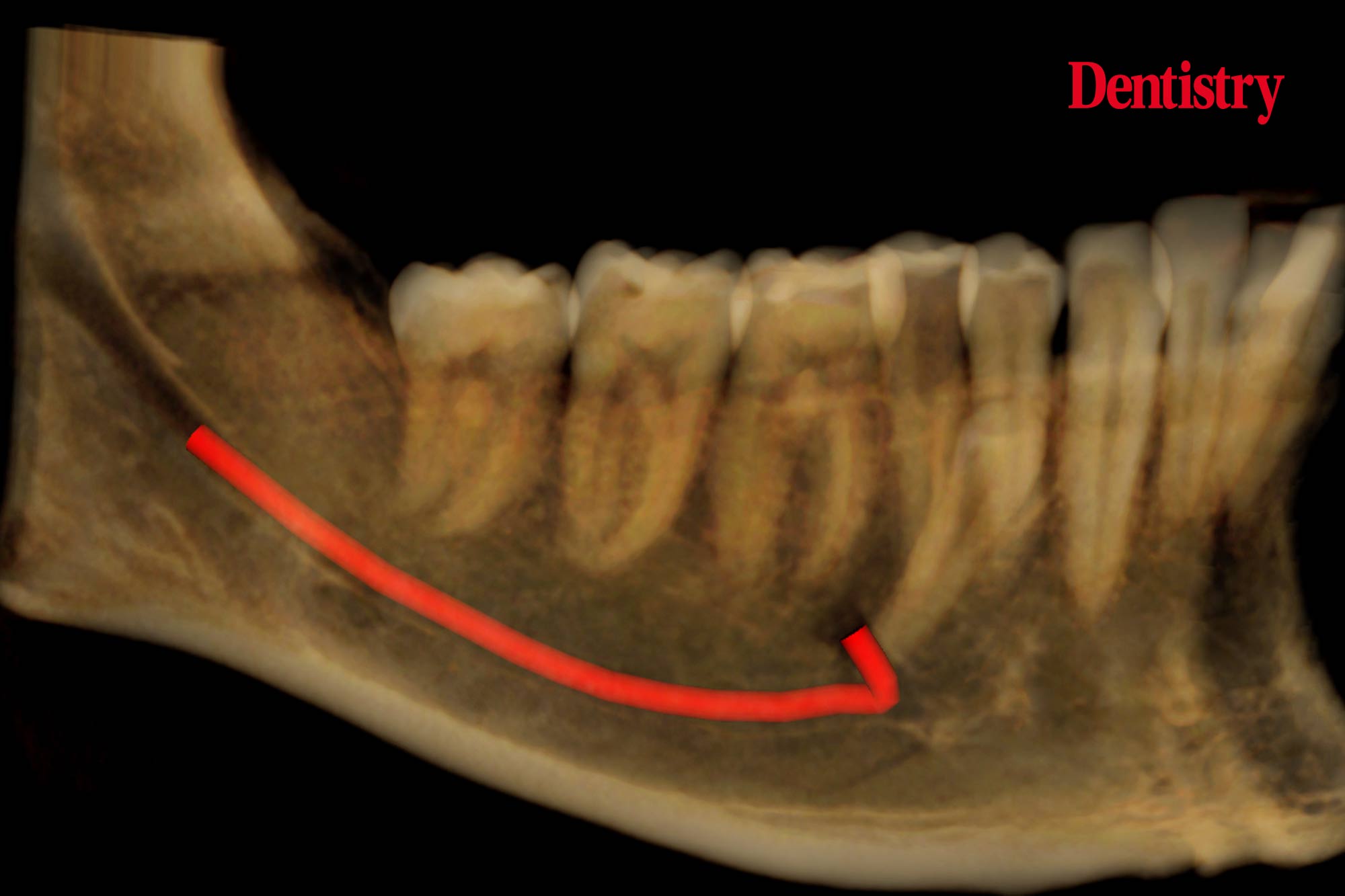
From a surgical perspective, the root of the supernumerary tooth is straight and its apex contacts the ID canal at the point at which the mental canal is given off (Figures 5 and 6). The only potential risk would be with downward pressure.
In a surgical context there is a potential risk with any applied downward pressure.

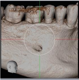
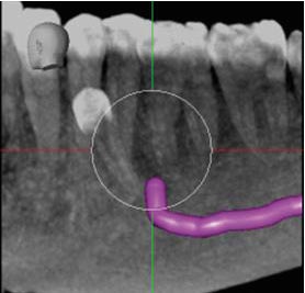
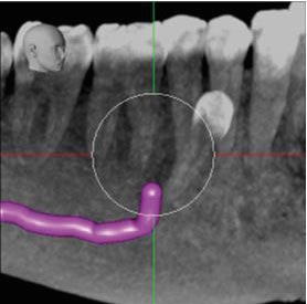
Volume-rendered image of buccal surface of mandible showing mental foramen position and two Maximum Intensity Projection images with the ID and mental canals marked.
For more information visit dental-scan.co.uk.

