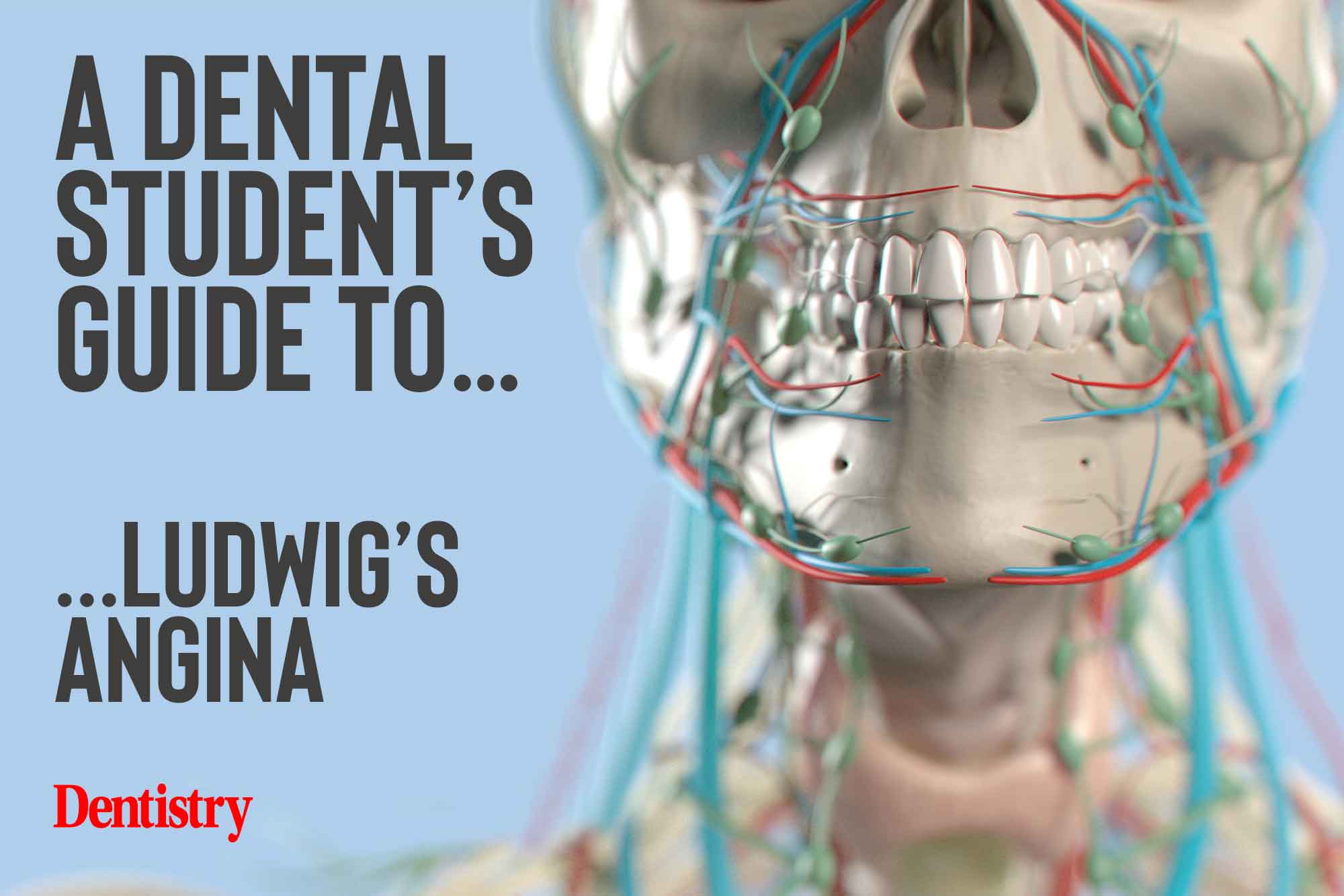
 To kick start this year’s dental student guides, Hannah Hook covers Ludwig’s angina – its causes, symptoms and treatments.
What is Ludwig’s angina?
Ludwig’s angina is a medical emergency caused by bilateral cellulitis of the submandibular, sublingual and submental spaces (Main et al, 2016).
It most commonly arises from a lower molar tooth whose roots lie below the level of the mylohyoid ridge. This enables infection to penetrate below the level of the mylohyoid muscle and easily cross the midline. It rapidly spreads to involve submental and sublingual spaces.
Mandibular second and third molars are accountable for approximately 90% of cases of Ludwig’s angina. However, other causes such as laceration, mandible fracture, osteomyelitis and sialolithiasis have been identified.
Whilst Ludwig’s angina often develops in healthy patients, predisposing factors include malnutrition, immunosuppression, HIV, diabetes, alcoholism and oral malignancy.
Ludwig’s angina results in obstruction of the airway and if not quickly treated is life threatening.
How does it occur?
As stated above, Ludwig’s angina most commonly originates from a dental infection in the second or third mandibular molars.
The infection progresses through the tooth or the subgingival pocket. It passes inferior to the mylohyoid ridge below the mylohyoid muscle into the submandibular space. It then rapidly progresses to involve the sublingual and submental spaces and crossing the midline.
The infection can also track posteriorly and involve the retropharyngeal and pharyngomaxillary, encircling the airway (Candamourty et al, 2012).
Onset of the swelling is usually acute. And due to the rapid progression of the condition, immediate treatment to avoid asphyxiation is essential.
Signs and symptoms
- Pain
- Fever
- Dysphagia
- Drooling – due to inability to swallow, including patient’s own saliva
- Hot potato or hoarse voice
- Difficulty breathing.
Extra-orally:
- Bilateral swelling of the neck
- Swelling is firm
- Unable to palpate lower border of mandible
- Restricted mouth opening.
Intraorally
- Raised and firm floor of mouth
- Restricted tongue movements.
Treatment
If Ludwig’s angina is identified in general dental practice, the patient should be immediately sent to the nearest hospital for treatment.
Once in the hospital department, healthcare staff will assess the patient’s airway and manage it appropriately.
The patient will receive intravenous antibiotics for the infection and steroids to reduce swelling and pain relief. Blood tests will help measure the inflammatory marker C-reactive protein. And clinicians will also assess white blood cell levels.
The patient will then undergo a general anaesthetic for emergency surgery.
The causative tooth is extracted, the infected spaces explored, and the infection drained. This usually requires both an intra and extra-oral approach.
Intraorally, if no pus drains from the socket a flap will be raised, any pus encouraged to come out, and also a drain is placed.
Extra-orally the clinician will make an incisione 2-2.5cm below the lower border of the mandible. They will then explore the space, drain the pus drained, and place a drain. The placement of drains helps to keep the spaces open and prevents pus forming a collection (Choi, 2017).
Key points
- Ludwig’s angina is a medical emergency that requires immediate treatment
- It most commonly occurs due to infection of the second or third mandibular molars
- Symptoms include severe swelling of the submandibular region, raised floor of mouth, reduced mouth opening and difficulty swallowing, breathing and opening their mouth
- Patients require admission to hospital for IV antibiotics, steroids, pain relief and emergency surgery.
Catch up with previous student’s guides:
Follow Dentistry.co.uk on Instagram to keep up with all the latest dental news and trends.
References
Candamourty R, Venkatachalam S, Ramesh Babu MR and Kumar GS (2012) Ludwig’s angina – An emergency: A case report with literature review. J Nat Sci Biol Med 3: 206-8
Choi MG (2017) Modified drainage of submasseteric space abscess. J Korean Assoc Oral Maxillofac Surg 43: 197-203
Main B, Collin J, Coyle M, Hughes C and Thomas S (2016) A guide to deep neck space fascial infections for the dental team. Dent Update 43: 745-52


