
How would you improve the appearance of a single dark incisor? Shiraz Khan shares his award-winning tooth whitening case from the Dentistry Clinical Case Awards 2022.
As has been reported on many occasions, the ability to improve a patient’s smile can have profound effects on their confidence. This subsequently can also lead to improvements in oral health (Baldwin, 1980; Davis, Ashworth and Spriggs, 1998).
Regarding noticeable tooth discolouration, some have considered this to be a physical handicap. It can impact a ‘person’s self-image, attractiveness and even employability’ (Kelleher and Roe, 1999).
The single dark central incisor poses many challenges aesthetically and restoratively. To avoid excess tooth preparation to create restorative volume and mask the tooth, the substrate should undergo tooth bleaching. This attempts to reduce the prominence of any tooth discolouration irrespective of the definitive treatment plan.
This upholds the principles as outlined by Banerjee (2013) regarding minimally invasive dentistry, by minimising the amount of tooth preparation required to achieve a functional and aesthetic result.
Simple, staple, safe
Home whitening is considered a simple, staple and safe aesthetic treatment. It can be carried out in the absence of the any dental disease (Kelleher and Roe, 1999). This implies that prior to any whitening, or aesthetic treatment, the patient should be considered to be in stable oral health. Namely, free from any dental, periodontal or soft tissue disease.
Non-vital tooth bleaching has been shown to improve the appearance of the dark, discoloured tooth. Poyser, Kelleher and Briggs (2004) discuss the traditional method where an access cavity is created removing any gutta percha until the CEJ, sealing the root treatment and subsequently placing a cotton wool pledget with 10% carbamide peroxide, which is changed two-hourly and left in overnight, while bleaching externally.
More recently, a novel method of internal and external tooth whitening was reviewed and discussed by Greenwall-Cohen and Greenwall (2019), which is known as the ‘inside-outside closed’ (IOC) technique.
This process describes appropriate assessment and diagnosis that an existing root treatment is of a good standard. Following this, the cavity is appropriately debrided to ensure removal of any pulp-horn remnants and previous restoratives. The choice can be either 10% or 16% carbamide peroxide, which is sealed into the access cavity with a temporary restoration. The tooth (discoloured only) is then bleached externally to refresh the peroxide from internally as well as having an external effect.
The significant advantage of this technique rather than the previous described ‘open technique’ is that the patient is not having to place bleach into the cavity and keep refreshing this every two hours.
The process certainly takes more time, however, it offers the clinician greater control on the whitening outcome.
Case presentation
A 27-year-old female, who worked in the financial sector, attended the practice for a new patient assessment.
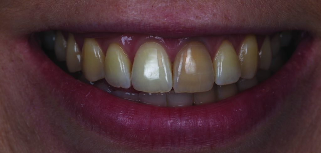
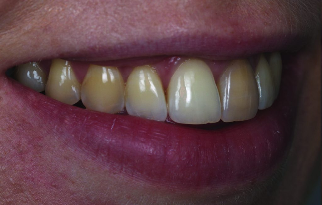
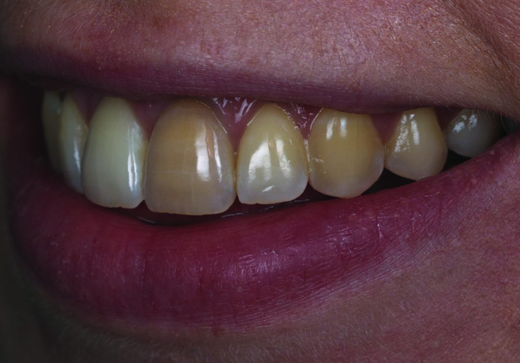
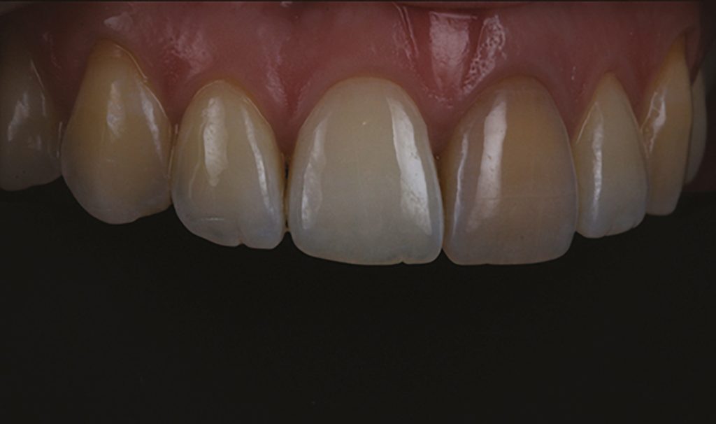
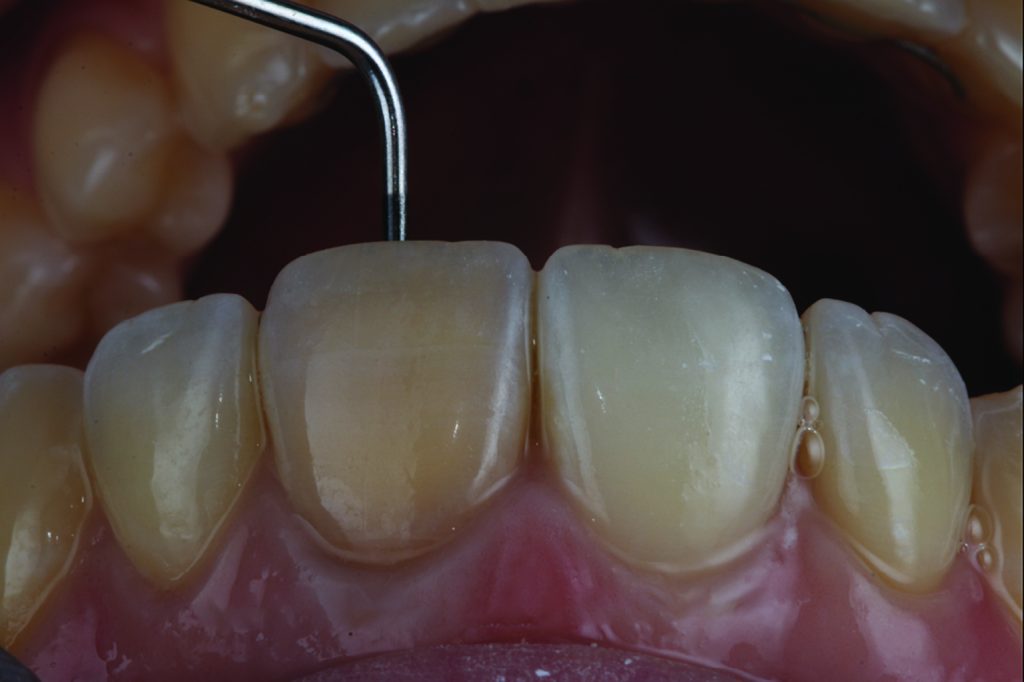
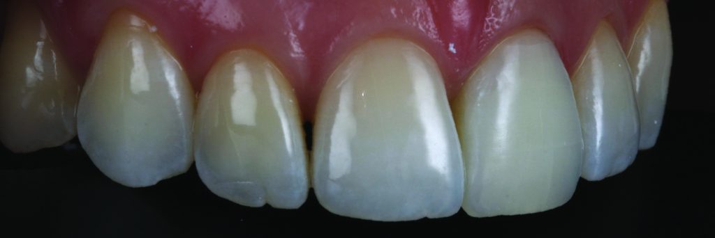
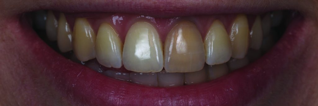
She presented with no pain and a single tooth veneer on her upper left central tooth to mask the discolouration (Figures 1 to 5). Medically, she was fit and well, with no allergies or routine medications disclosed. She was brushing twice daily with an electric toothbrush and flossing once weekly. Her previous dentist appointment was 12-months ago and attends hygiene appointments bi-annually.
On examination, the patient presented with generalised gingivitis and a largely discoloured UL1. The tooth had trauma some 10-years ago while playing hockey. The tooth had been root treated and had no pain or symptoms since the root treatment. The root filling was completed to a high standard with good length compaction, and completely free of any periapical or peri-radicular widening of the lamina dura or radiolucency indicating re-infection.
Palatally, the tooth was well sealed with composite (shade A3), which demonstrated no signs of leakage or secondary caries. Otherwise, the patient was caries free, as confirmed by radiographic examination.
The patient was diagnosed with generalised gingivitis and mesiopalatal. Aesthetically, she was diagnosed with generalised extrinsic staining with severe discolouration of the UL1.
Discussion of the patient options
The patient was keen to have a veneer restoration to mask the UL1.
As we are aware, the single central incisor is the most difficult tooth to replicate aesthetically from its contralateral counterpart. This is without the excessive discolouration this case presents.
As aforementioned, the restorative volume required to create this masking effect will lead to somewhat over-preparation of the teeth. Therefore, even if a restorative approach was to be considered, preoperative whitening would be a staple requirement.
My discussion with the patient was straight-forward and simple. If the restorative option was to be considered, I would only advise this to be considered once the whitening had been completed.
The patient was grateful for the openness of the discussions and agreed to undertake non-vital ‘inside-outside closed’ (IOC) whitening.
Creating access and beginning whitening
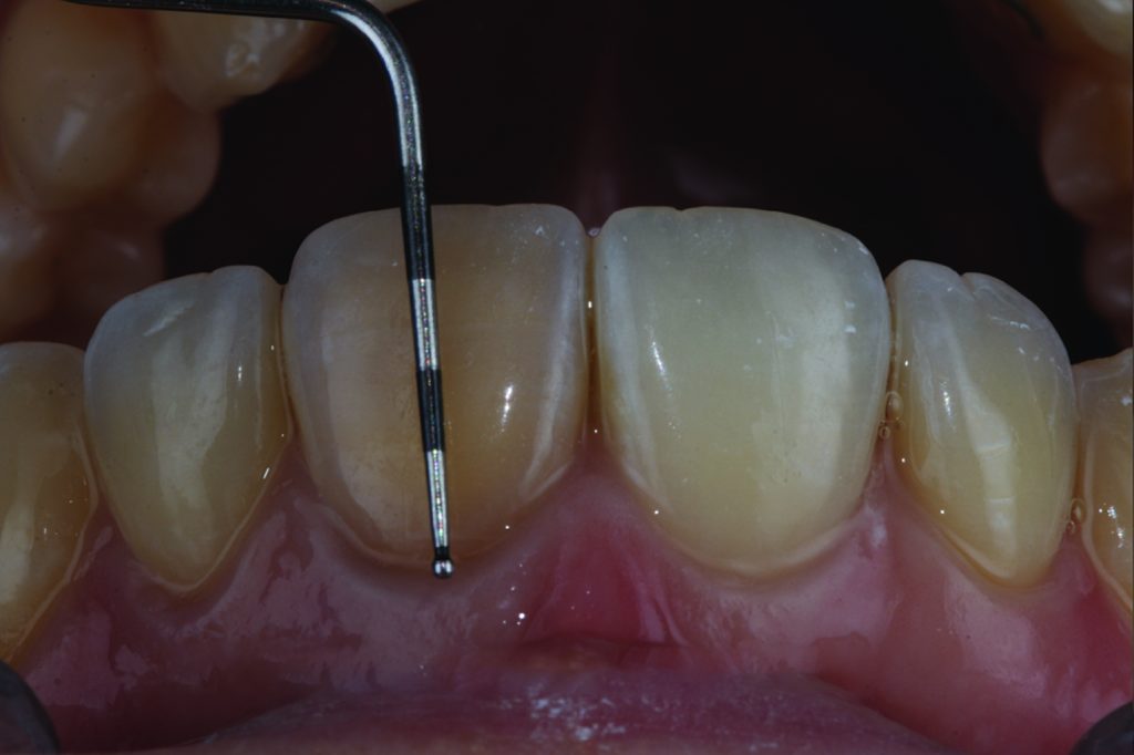
A full mouth supra and subgingival scale and polish was completed with the hygienist.
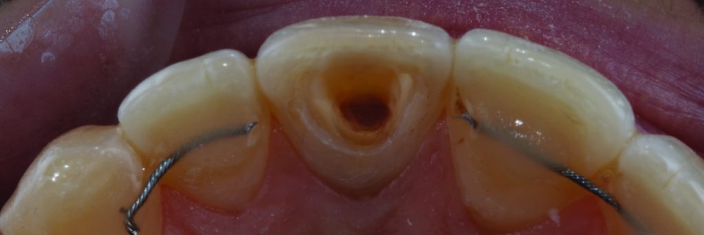
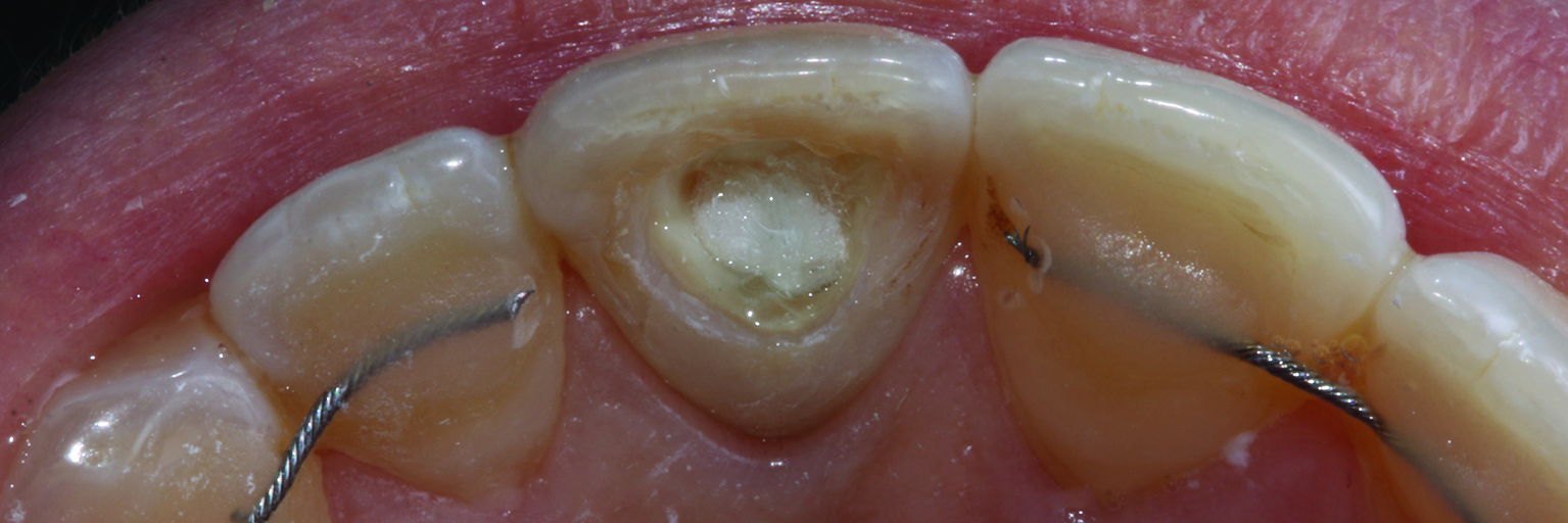
The patient attended for an appointment for the retainer to be removed and access to be created (Figure 6), the root treatment was already sealed with a restorative, and the 16% carbamide peroxide was sealed with cotton pledget and was temporised (Figure 8).
The patient then undertook two weeks home whitening with 16% carbamide peroxide on the front tooth only. This was reviewed after two weeks and repeated for two further cycles.
Bond strength can reduce by up to 20% if completed on the same day as the cessation of the whitening (Garcia-Godoy et al, 1993; Nour El-din et al, 2006). This occurs due to thin sparse and fragile resin-tag formation. A period of two weeks allows for any hydroxyl radicals to leach out of the teeth, and for the whitening result to stabilise. Therefore, the appointment for definitive palatal composite of the UL1 was to be completed two weeks after completion of internal and external whitening.
Etching and finishing
The tooth was etched with 37% phosphoric acid for 30 seconds (enamel) and 15 seconds (dentine) in case of UR1 MP. The teeth were washed thoroughly for 30 seconds and then air dried. The bonding system used was Optibond FL and cured for 30 seconds on each tooth.
We opted to use Venus Pearl composite system in this case. We chose shade BXL, as the previous composite used was a chromatic A3 shade. The intention behind this was for brightness of the core composite to shine through to the tooth substrate. This would also manage the shade rebound that would be likely to occur post-whitening. The final cure was completed under glycerine gel and the finishing protocol began. The palatal surface is polished with Astropol finishers (grey, yellow and pink) followed by the 3M enhanced polishing spirals.
A smooth surface palatally is not of any aesthetic advance but rather for patient comfort and reduction in stain retention. Restorations of this nature have been shown to be successful in 97.6% of cases after 10 years (Opdam et al, 2014).
Finally, impressions were taken for a new fixed retainer, which was subsequently bonded in place from upper canine to canine.
Conclusion and reflection
The discussion process and valid informed consent are essential when undertaking all forms of treatment, but particularly with reference to elective or aesthetic treatment.
The discussion process with this patient was very important, as she was set on undertaking a restoratively led treatment plan. While there is no doubt a satisfactory result could likely be achieved, this would be at the cost of significant over-preparation to mask the discoloured tooth, which may not have been required.
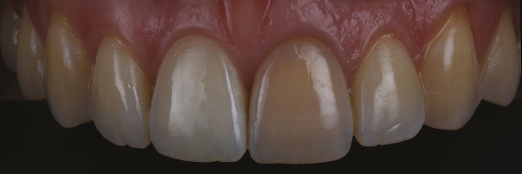
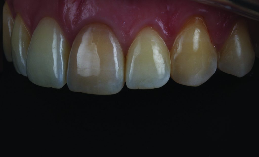
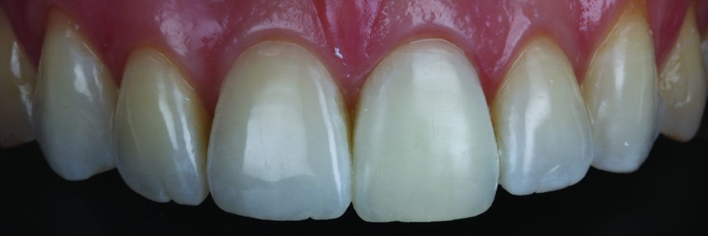
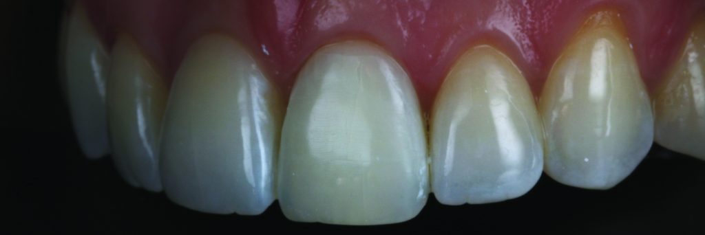
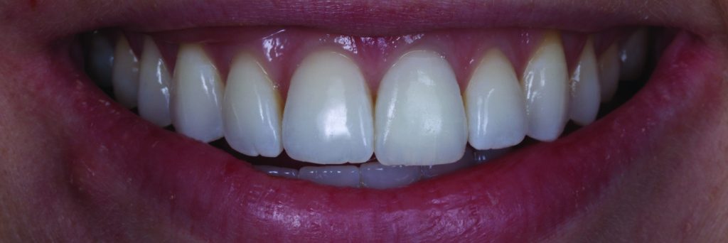
Figures 9 to 11 show the result of the whitening after four weeks. As demonstrated in this tooth whitening case, a simple yet refined intervention created a suitable outcome without the need for restorative intervention.
The patient was ecstatic in this case as she was previously informed that the only method to create a reasonable outcome would require preparation of the tooth for a veneer.
With consent being a dynamic process, there is no need to assume that the patient would not still consider additional treatment after the whitening had taken place, but discussion of the options at each stage of treatment and appropriate documentation are imperative elements to informed consent.
The patient was delighted by the restorations completed and enhancement to her smile.
To improve the case, I would perhaps encourage the patient to whiten her remaining teeth for a week with 16% carbamide peroxide. In addition, I would have made the patient a removable retainer during the process to avoid any unwanted tooth movement.
Read more National Teeth Whitening Day articles:
- How to achieve simultaneous straightening and whitening
- See the Light with Pola Light professional whitening
- Tooth whitening as an entry point to better oral health
- How is the demographic for tooth whitening changing?
Follow Dentistry.co.uk on Instagram to keep up with all the latest dental news and trends.
For references, email rowan.thomas@fmc.co.uk.

