Alex Woodham discusses a case where CBCT is used to evaluate sinus anatomy before commencing dental implant treatment.
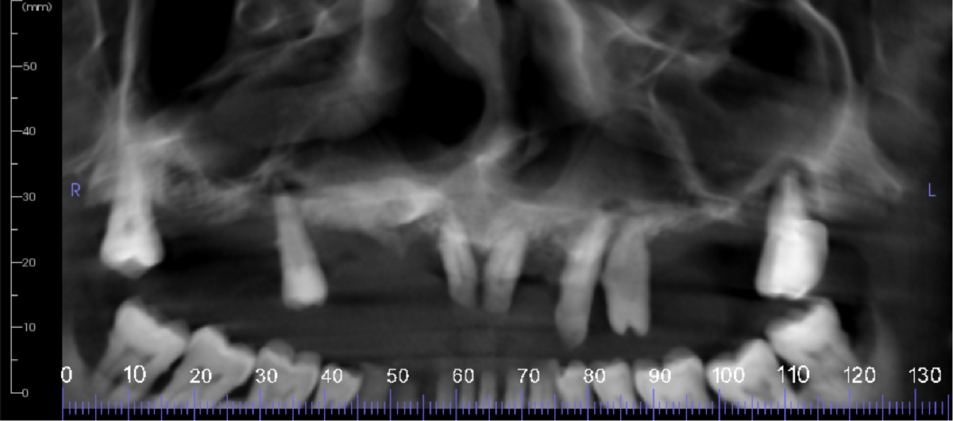
There is evidence of periapical pathosis on teeth UR5, UL3, UL4 and UL7:
- The UR5 has severe periodontal loss of attachment and an apical radiolucency with lifting and erosion of the nasal floor
- The UL3 is root filled, but just short of the apex with an apical radiolucency also involving the UL4
- The UL7 is root filled but short of the apices. There are radiolucencies on all roots with severe periodontal loss of attachment. The sinus floor shows erosion.
The UR3 is unerupted, lying horizontally touching the floor of the right side of the nose. The apex is touching the root of UR5 and incisal tip close to the UR1 apex. There is external resorption of the buccal side of UR3 crown possibly involving the pulp.
There is chronic bilateral maxillary sinusitis. Both sinus ostia are full of soft tissue and appear to be widened. There is no displacement or erosion of the sinus walls.
A differential diagnosis of this appearance is an inverted papilloma of the right ostia and sinus cavity. Further investigation is necessary to confirm a diagnosis.
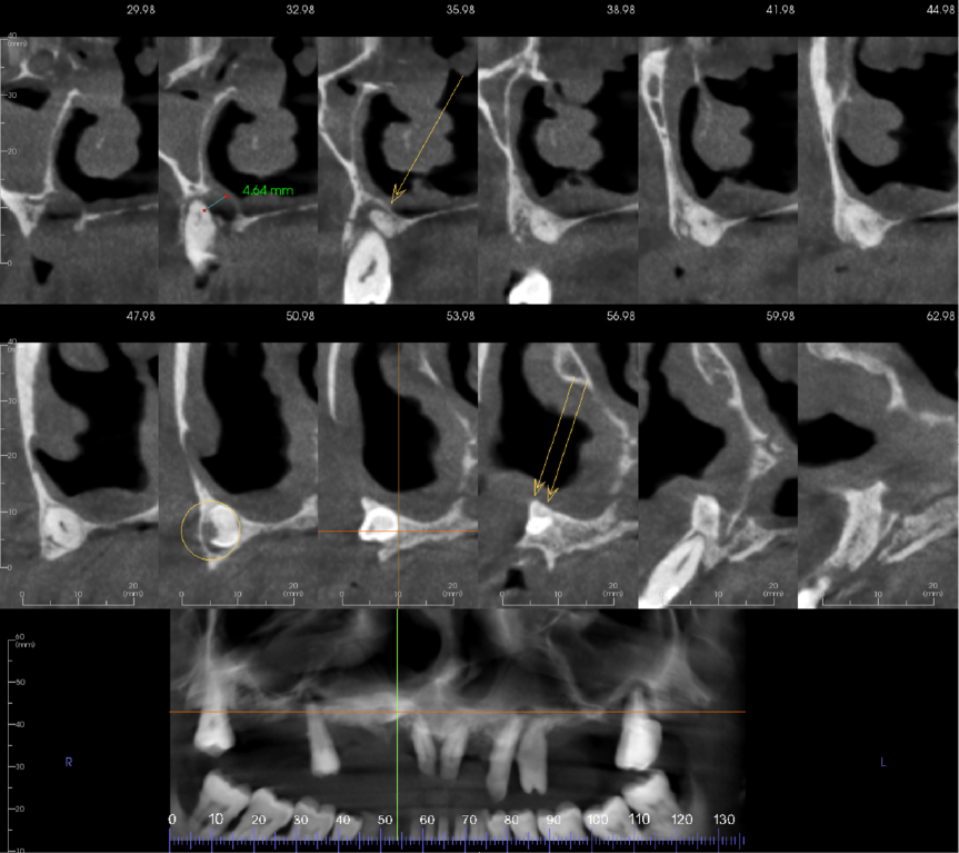
Cropped panoramic and 12 cross sections with 3mm spacing between slices of alveolar ridge showing the unerupted UR3 and its relationship with the UR5 and UR1 (figure 2).
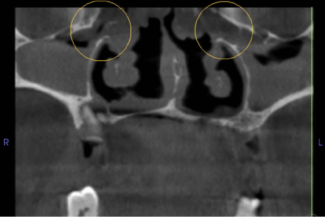
Coronal section through both maxillary sinus ostia (circles in figure 3). Both sinus ostia are full of soft tissue and appear to be widened. The posterior portions of the sinuses are almost completely full of soft tissue although there is no displacement or erosion of the sinus walls.
Learning from this case
CBCT is commonly used to evaluate sinus anatomy before commencing dental implant treatment. Literature reports periodontal disease and periapical pathology increases the risk of maxillary sinusitis.
Sino-nasal inverted papilloma is a benign but locally aggressive tumour. It usually presents in patients with a history of sino-nasal infections.
For more information visit dental-scan.co.uk.

