In the latest case of the week, Alex Woodham discusses how a CBCT was needed to check for apical pathology before root canal treatment.
LR6 has a history of pain and tenderness to biting for two years. The patient had symptoms which correlate to an irreversible pulpitis following the replacement of the restoration, which resolved with antibiotics. Clinically the tooth is not TTP, with no tenderness in the buccal sulcus and probing depths within normal limits. CBCT needed to check for any apical pathology before commencing root canal treatment.
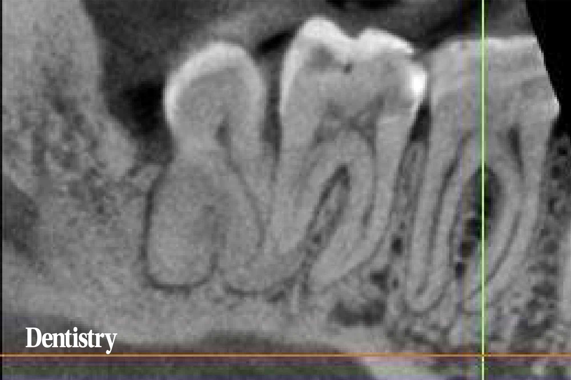
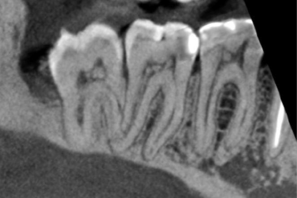
The LR6 has patent root canals, normal periodontal ligament width and lamina dura. The most likely explanation for the pain is a non-vital pulp and/or a root fracture. No root fracture is visible.
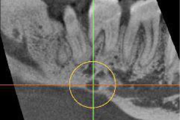
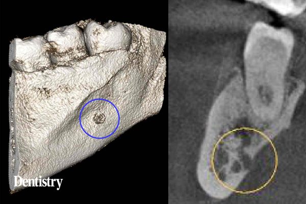
In the lingual cortical plate below the LR7 is a well-defined unilocular radiolucency, 4mm wide with loss of the external surface of the cortical plate and continuous with the bone marrow space. It is very close to the mandibular canal and its inflammation could cause pain.
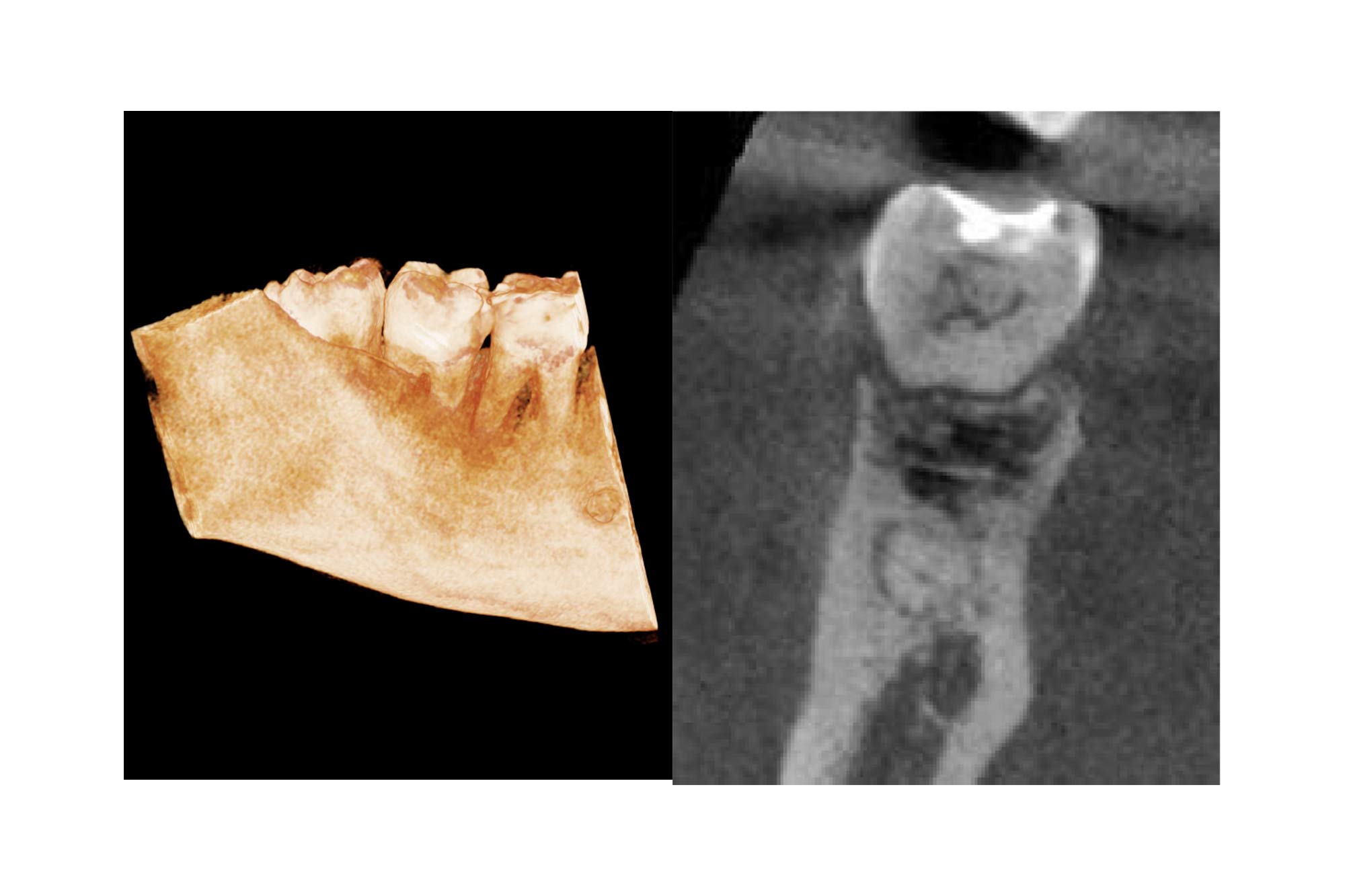
The LR8 is impacted, the apices are not close to the mandibular canal. There is an enlarged follicle space, which does not have the typical appearance of an odontogenic cyst and is also likely to be from past episodes of pericoronitis.
The LR6-8 have pulp stones.
Learning from this case
A root fracture might be suspected, but will not always be visualised in a CBCT scan if it is smaller than the smallest voxel size available when taking the scan.
Rarely, a finding such as the radiolucency in the LR7 region could be a serious lesion such as a multiple myeloma and a further follow up would be recommended with an oral surgeon.
Pulp stones are foci of calcification in the dental pulp of the tooth. When doing root canal treatment their presence can make the location of root canal orifices difficult.
For more information visit dental-scan.co.uk.

