Katarzyna Ostrowska shares an initial caries management case in which guided enamel regeneration was used to arrest the development of a carious lesion.
Caries treatment is often equated with the treatment of cavitated lesions that necessitate a restorative approach. However, the initial, non-cavitated stages of caries, when the lesions are still in the enamel, are often left untreated the so-called ‘wait and watch’ approach (Shah et al, 2023). This is because, on the one hand, there is a lack of tailored solutions for such incipient lesions. On the other, a restoration would lead to the sacrifice of a significant amount of healthy tooth structure.
The self-assembling peptide technology, incorporated in Curodont Repair, and working through ‘guided enamel regeneration’ fills the gap between preventive and restorative strategies. It is based on a short, ‘intelligent’ peptide, P11-4, which self-assembles into a three-dimensional biomimetic matrix within the subsurface body of initial carious lesions.
This matrix serves as a platform for calcium and phosphate ions from patients’ own saliva for in-depth de novo hydroxyapatite formation over the next weeks and months, mimicking the natural biomineralization process (Kind et al, 2017).
Curodont Repair is a patented, fluoride-free formulation that is applied in a short in-office procedure that can be done by dentists or hygienists. In the majority of cases, only one application is required for treating a lesion. The procedure is non-invasive, pain-free, and non-staining.
The efficacy of Curodont Repair has been proven in several studies, including randomised controlled clinical trials. It enables not just a significantly superior rate of caries arrest than fluorides but in fact caries regression through in-depth enamel regeneration (Bröseler et al, 2020; Welk et al, 2020; Godenzi et al, 2023; Alkilzy et al, 2018).
Case report
Chief complaint
An 18-year-old male patient arrived with the chief complaint of multiple white discolourations on his front teeth, which led to embarrassment in social interactions. The patient gave a history of undergoing fixed orthodontic therapy during which he experienced difficulty in oral hygiene maintenance.
Intraoral examination
White discolourations of varying sizes were noted along the gingival margins of multiple maxillary and mandibular anterior teeth. These areas, which had rough surfaces and were chalky in appearance, were visible both when the surfaces were wet and dry. In particular, the maxillary left central incisor (21) presented a large white discolouration covering almost the entire cervical third of the labial surface of the crown.
Diagnosis
Early-stage caries (ICDAS Score 2), as a result of improper oral hygiene maintenance during the fixed orthodontic therapy, were diagnosed, including for 21.
Treatment plan
Taking into account the oral findings, the patient’s young age and his unsatisfactory experience with the past dental treatment, a decision was made to treat the lesions with Curodont Repair in a non-invasive manner and without pain.
Treatment procedure
Thorough oral prophylaxis was performed to eliminate all deposits. Next, the affected teeth cleaned with 2% sodium hypochlorite saturated in a cotton pellet. The teeth were then rinsed and dried. Next, 35% phosphoric acid was used to etch the lesion surfaces for 20 seconds, followed by rinsing. The treatment site was isolated using cotton rolls and dried gently.
One applicator of Curodont Repair was activated and the saturated sponge was squeezed well on the surface of the lesion.
After a waiting period of five minutes, the patient was discharged with routine oral hygiene instructions.
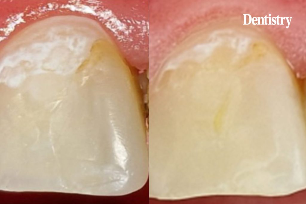
Follow up
Within two months of the treatment, the early carious lesion demonstrated a significant reduction in size and appearance, indicating caries regression.
vVARDIS is a new exhibitor at British Dental Conference & Dentistry Show Birmingham! For more information, visit vVARDIS at booth F64.
References
- Shah SV et al. J Am Dent Assoc. 2023;154:897-909.e6.
- Kind L et al. J Dent Res 2017; 96:790-797
- Bröseler F et al. Clin Oral Investig. 2020; 24:123-132
- Welk A et al. Sci Rep 2020; 10:6819
- Godenzi D et al. J Am Dent Assoc. 2023:S0002-8177(23)00416-6
- Alkilzy M et al. J Dent Res 2018; 97:148
This article is sponsored by vVARDIS.

