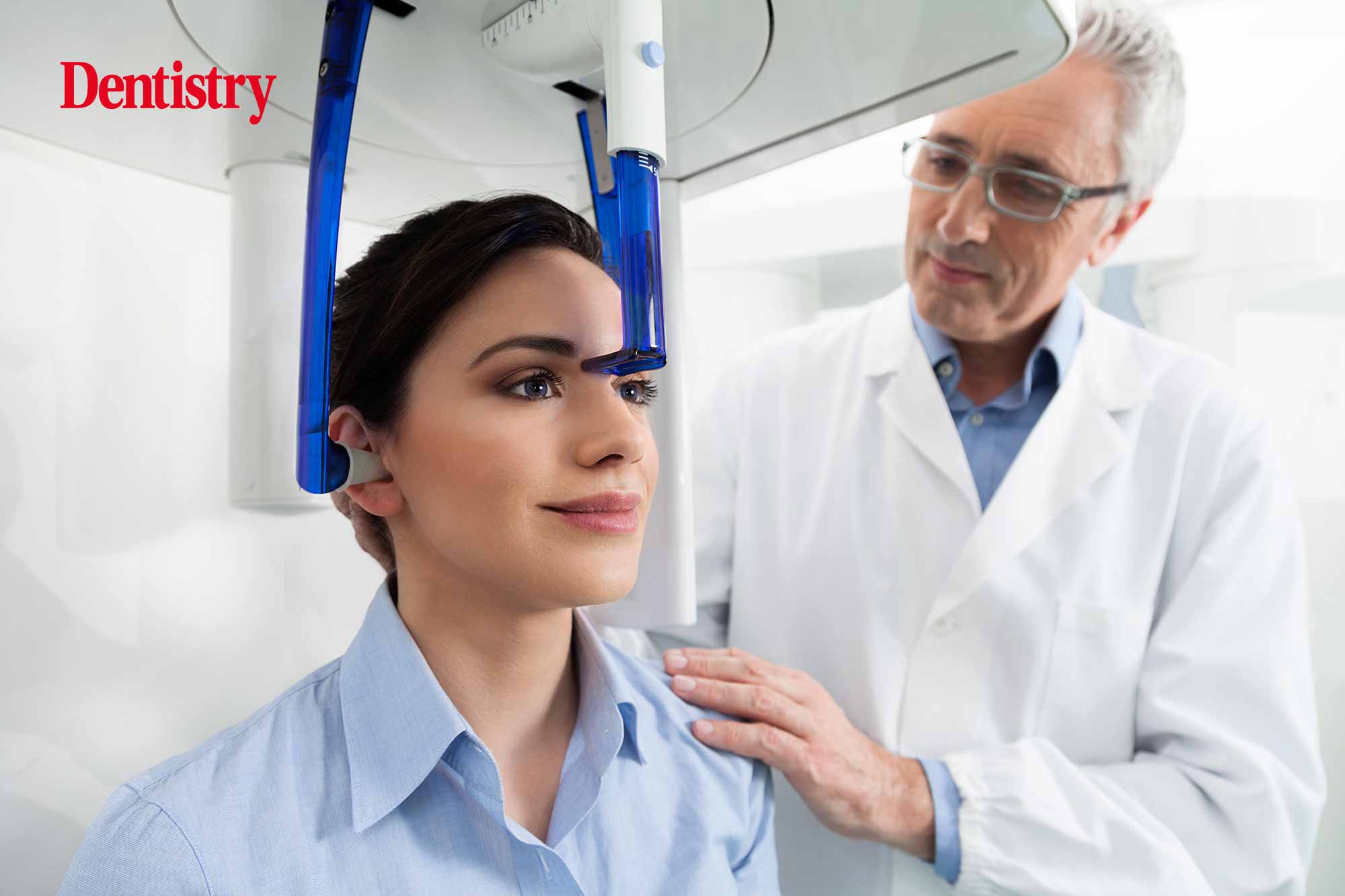
 Artur Silva considers the benefits of investing in CBCT and what to look for when making buying decisions.
Cone beam computed tomography (CBCT) entered the field of dentistry in the late 1990s/early 2000s. It resulted in a step-change in the way dentists can view their patients’ mouths.
There is growing evidence that patients and dental professionals have benefitted considerably as a result of using CBCT. Both to plan treatment and for postoperative evaluation.
Some suggest that, for certain cases, using CBCT is compulsory. The level of information the dentist receives from this technology serves to reduce the possibility of complications. As well as resulting in reliable, successful treatment outcomes.
These benefits include:
- Detailed surgical/implant planning and execution of the desired treatment
- Operational predictability (aided by surgical guides, for example)
- Use of the correct volume and type of augmentation material
- Minimisation of post-operative complications by using novel equipment such as virtual endoscopy to trace crucial anatomical structures.
All this, in turn, outweighs the risks associated with exposing the patient to a CBCT dose.
Choosing and using CBCT
So, as we know, CBCT can help plan a case. However, planning for CBCT itself can also require some planning!
Overcome this challenge by making sure you choose your CBCT carefully. So that it, for instance, has easy-to-follow icons in the operating software, guiding the practitioner through a series of settings to choose.
The ideal CBCT unit should also incorporate many different clinical applications; such as panoramic and cephalometric, in addition to 3D imaging of very small to larger fields of view.
Furthermore, as touched upon above, of incredible importance is having a machine that will target a focus area with the correct level exposure. This way the patient is exposed to no more radiation than is necessary.
As Bornstein and colleagues wrote in 2014: ‘Practitioners who prescribe or use CBCT units should design specific CBCT equipment protocols that are task specific and incorporate the imaging goal for the patient’s specific presenting circumstances.
‘The protocol should include considerations of exposure (mA and kVp), minimum image-quality parameters (eg, number of basis images, resolution), and restriction of the FOV to visualise adequately the region of interest.’
After these protocols are established and the risks versus gains are determined, the clinician will have a better understanding of when to use CBCT. Especially when the benefits are clear for both the practitioner and the patient.
Interestingly, once the decision has been made for a particular patient to have a CBCT image taken, it can then also play a significant educational role. For example, the image can show the patient the case in question. As well as the pathology and related anatomy, and any specifics of the treatment.
Patients value helpful visual, evidence-based information.
Picture the future with CBCT
Patient demand for highly aesthetic outcomes is higher than it has ever been and will not diminish.
To meet patient expectations, dentists must make use of the highly sophisticated and advanced technologies that are available today. Most especially in the fields of implant therapy, periodontal treatment, endodontics, and restorative care.
Clearly, the more information the clinician can gather in advance of treatment, the better the planning and outcome.
To that end, 3D imaging techniques such as CBCT can play a pivotal role in providing the clinician with the tools for planning a more predictable case. This will therefore minimise the risk of possible complications, and facilitating improved outcomes.
Having a 3D imaging system adds true value to the dental practice for both patients and the dental team. Something that we at Hague Dental recognised some years ago.
Over time, exciting developments in dental diagnostic technology has seen CBCT and 3D imaging systems become more commonplace in practices. If you are looking to join the 3D revolution, we can help you explore options considering low dose, image accuracy and detail and ease-of-use.
Simply visit www.haguedental.com, email [email protected] or call 0800 298 5003.
References
Bornstein MM, Scarfe WC, Vaughn VM and Jacobs R (2014) Cone Beam Computed Tomography in Implant Dentistry: A Systematic Review Focusing on Guidelines, Indications, and Radiation Dose Risks. Int J Oral Max Imp 29 (SUPPL): 55-77


