CBCT scanner: I-CAT Classic
CBCT imaging protocol: 60x80x80mm, 0.2 voxel
Effective dose: 0.03 mSv
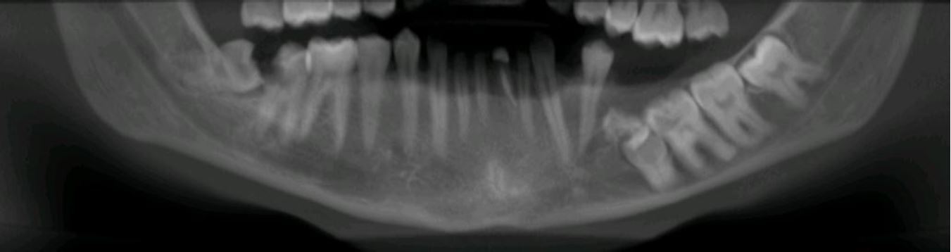
Clinical information: assessment of location of premolars and molars and relationship with IDN.
Radiographic impression
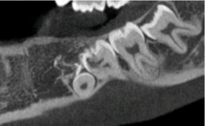
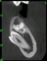
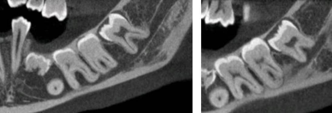
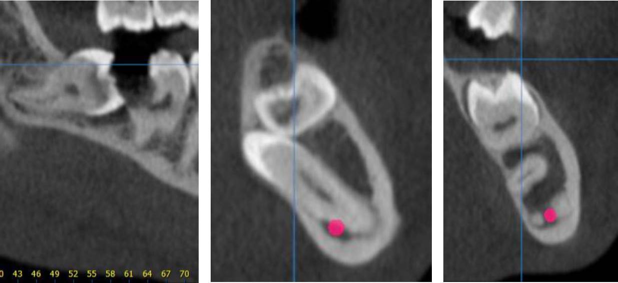
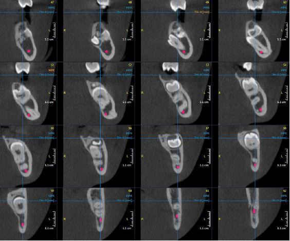
Mandible: the right mandibular premolars and molars are impacted. The primary second molar is ankylosed and impacted; it seems to be preventing the eruption of LL5 and directing it in a lingual position. The mesial aspect of the crown of LL6 is in contact with the impacted primary tooth and presents with an external resorption.
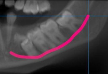
LL6, LL7 and LL8 present with apical dilaceration, the apices of LL8 are situated within the mandibular canal.
Impressions and recommendations
Ankylosed primary tooth with impaction of LL5, LL6, LL7 and LL8.
LL5 is in a lingual position, LL6-LL8 are impacted and their roots are dilacerated.
LR8 impacted in an oblique direction with large carious lesion on LR7.
For more information visit ct-dent.co.uk.