Basic report for pathology only
CBCT scanner: Kavo OP 3D Vision V17
CBCT imaging protocol: 16cm diameter x 10cm height
Effective dose: 0.1 mSv
Clinical information: assessment of both jaws prior to implant planning.
Click here to view and manipulate this case of the month CBCT on CT Dent’s Cloud Viewer.
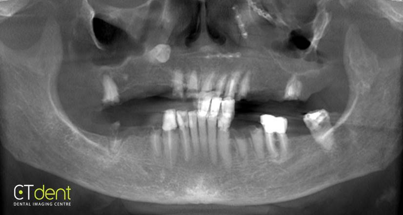
Click here to view and manipulate this case of the month OPG on CT Dent’s Cloud Viewer.
Findings
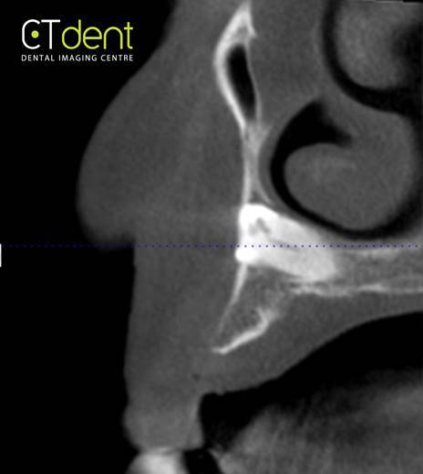
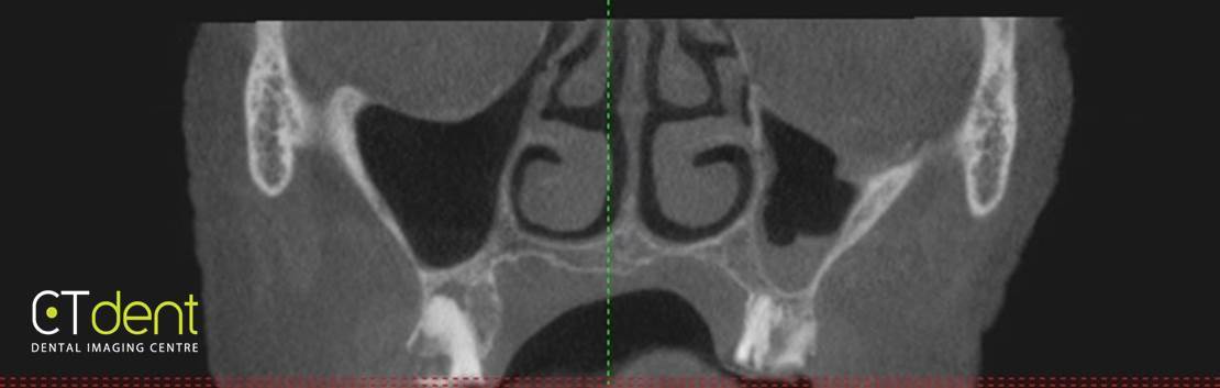
Paranasal sinuses: mild mucosal thickening with air bubble appearance in the left sinus
Nasal cavities: no abnormalities detected
Airway: no abnormalities detected
Osseous structures: no abnormalities detected.
Dental findings
Maxilla and mandible: presence of multiple residual root tips throughout the maxilla and mandible. UL3 is impacted in a high position with features of ankylosis and no associated pathology noted. Surgical plates and screws consistent with the history of trauma or orthognathic surgery.
Impressions and recommendations
The pertygomandibular ligament is calcified, surgical intervention is needed in presence of limited opening of the mandible.
Mild mucositis in the left sinus, no further evaluation needed. Carotid artery calcification: physical referral is suggested.
For more information visit ct-dent.co.uk.

