CT Dent introduces a case where its scanners were used to diagnose the patient.
Images provided
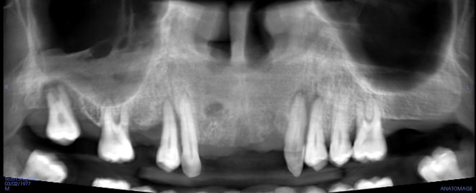
Cone beam CT images in the bone window. Axial, coronal and sagittal planes.
Clinical info
Implant analysis requested. Relevant history: please assess scan prior to implant placement.
Diagnostic objectives
- Implant planned
- Sinus evaluation
- Rule out pathology.
Findings
Maxilla: no abnormalities detected.
Sinuses: the right maxillary sinus is partially occupied by homogeneous area of increased density containing bubbles.
Nasal cavity: a deviation of the nasal septum was noted.
Other findings: no abnormalities detected.
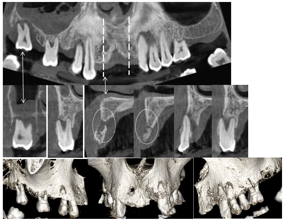
Dental findings: multiple cross-sections illustrating dental findings have been provided below.
Radiographic impression
Sinuses: the radiographic findings appear to be consistent with a chronic sinusitis of the right maxillary sinus. The presence of bubbles is a potential indication of an acute exacerbation of the chronic sinusitis. Review of the patient’s clinical history for chronic allergy or sinusitis is suggested; physician referral for more thorough evaluation is suggested if merited by clinical findings and symptoms.
Nasal cavity: deviation of the nasal septum is a common incidental radiographic finding and does not require treatment or referral unless the patient exhibits difficulty breathing through the nose.
Dental findings: please see the specific comments associated with each of the cross-sections illustrating dental findings below.
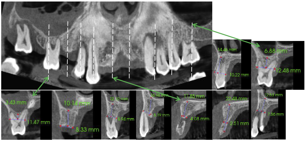
Implant measurements: in the absence of specific instructions related to areas of potential implant interest, the measured sites were arbitrarily selected to provide an indication of the bone available in various anatomic regions. Please make your own careful measurements once the number and specific location of each implant has been determined.
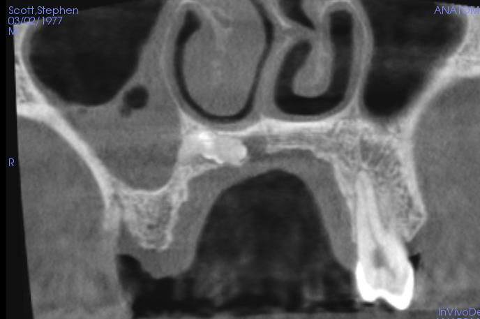
The following are selected images from the volume illustrating major findings
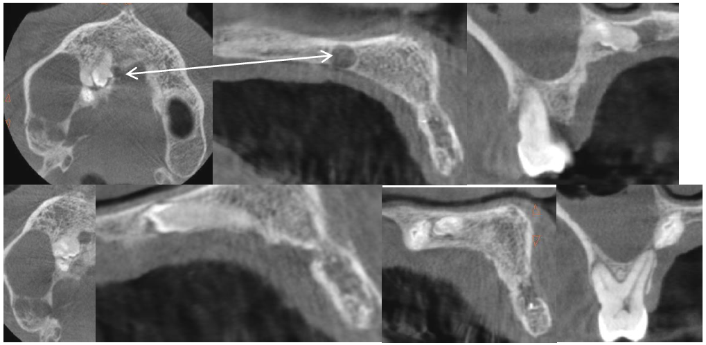
right maxilla extending from approximately the palatal surface of the lateral incisor posteriorly to the
palatal root of the first molar. A small well circumscribed corticated radiolucency was noted adjacent
to the tooth-like entity closest to the midline [arrows]. The radiographic appearance is consistent with
an odontoma (benign tumor of tooth forming tissue) with a small follicular/dentigerous cyst. The more
distal tooth-like object appears to extend beyond the palatal cortical plate adjacent to the apex of the
maxillary right first molar. Appropriate clinical evaluation and follow-up is deemed appropriate and is
suggested