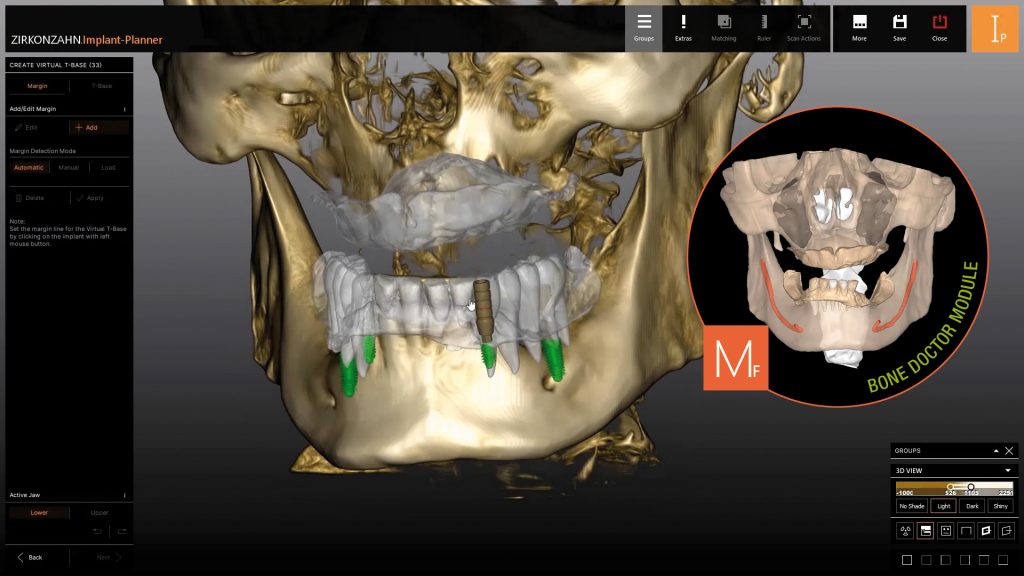
Zirkonzahn.Implant-Planner and Bone Doctor: implant planning software approved as a medical device and new software module for precise surgical planning.
With the Zirkonzahn.Implant-Planner software, the cooperation between dentists and dental laboratories can be taken to new levels, reconciling the planned aesthetic design of a prosthetic restoration with the planned implant situation (backward planning).
Based on digitally merged patient data (such as DICOM data, model scans, intraoral and facial scans), the dentist can determine the optimal implant position in terms of function and aesthetics, taking bone structure into account.
The software is available in two versions: Zirkonzahn.Implant-Planner as the full version for implant planning and the production of surgical guides, and Zirkonzahn.Implant-Planner Practice, which includes all essential functions exclusively for implant planning.
The user-friendly interface guides the dentist step by step through the entire planning process, enabling a straightforward data transfer to the dental lab. This allows the dentist to receive all components required for an implant case simultaneously (immediate loading). Production is carried out within the Zirkonzahn CAD/CAM system, from surgical guides to prosthetic restorations, or, thanks to the open data exchange function, also with CAD/CAM systems from other manufacturers or with 3D printers.
Bone Doctor software module
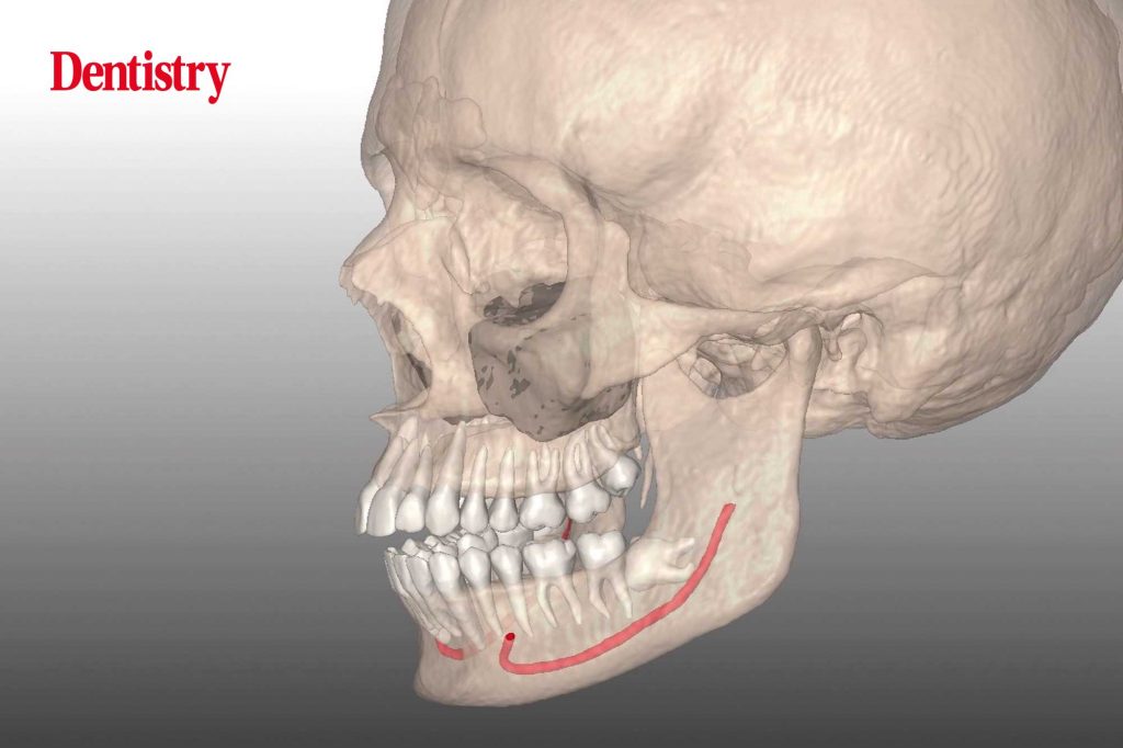
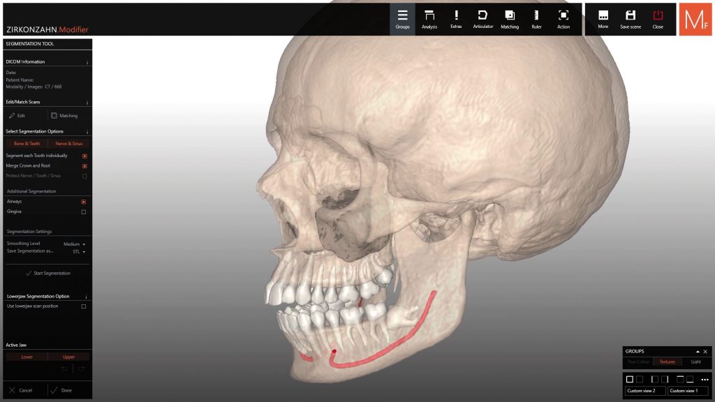
For even more precise implant planning, the 3D objects generated with the new Bone Doctor module of the Zirkonzahn.Modifier software can also be imported. This module significantly simplifies the digital analysis of the bone situation: by importing the patient’s DICOM data, the module allows users to analyse the different cranial bones and generate the corresponding 3D files.
The software is capable of autonomously segmenting the desired anatomical structures, such as the lower jaw, mandibular nerves, individual teeth, maxillary sinuses and other anatomical parts. Additionally, the extracted maxilla can be combined with the patient’s Real Movement data to analyse the condylar movements. Extracted teeth can also be used to perform orthodontic movements based on their actual root and crown morphology.

This article is sponsored by Zirkonzahn.

