This patient was sent to CT Dent for implant planning in multiple sites in the lower jaw.
CT Dent provides dental imaging for more than a thousand patients every month across seven dedicated imaging centres in the UK.
This imaging allows dental practitioners to make informed decisions for their patients, assisting with diagnosis and treatment.
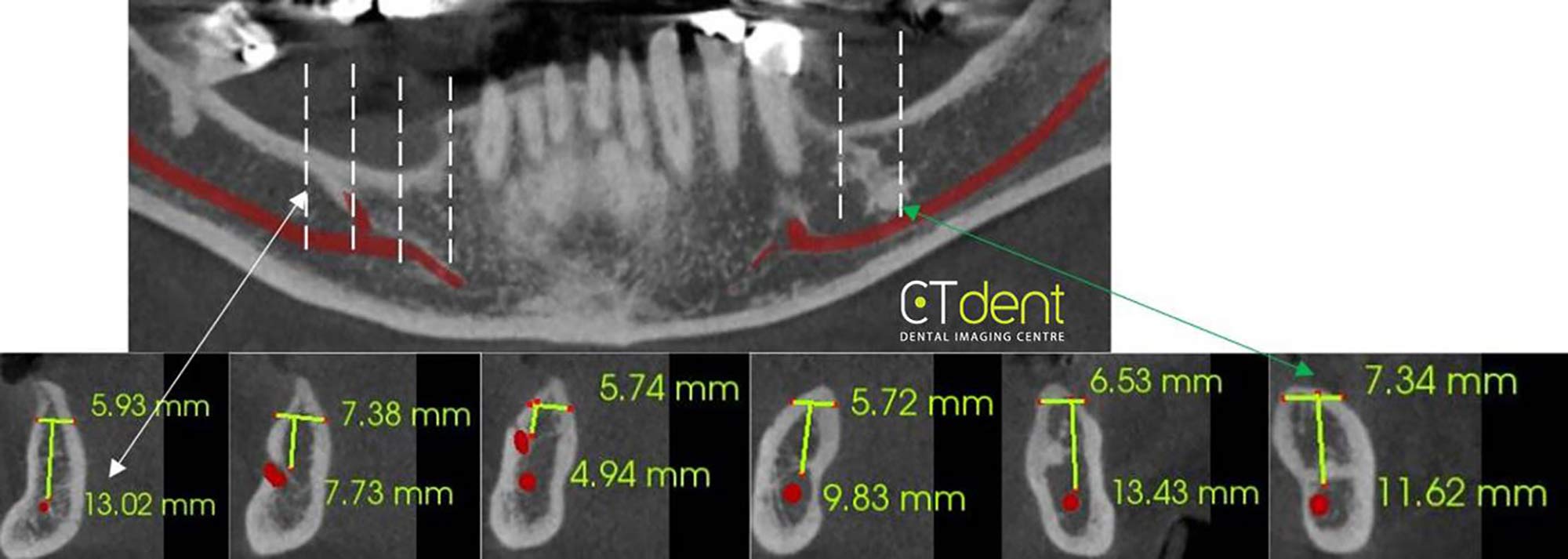
In this case, the use of cone beam computed tomography (CBCT) helped to plan for implants in the LL6, LR4 and LR6 sites in the lower jaw.
Uncommon
The Kavo OP 3D Vision (V17 model) took the scan. The field of view used 6cm height x 16cm width, with a resolution of 0.25 voxels. The effective dose to the patient was 0.12mSv.
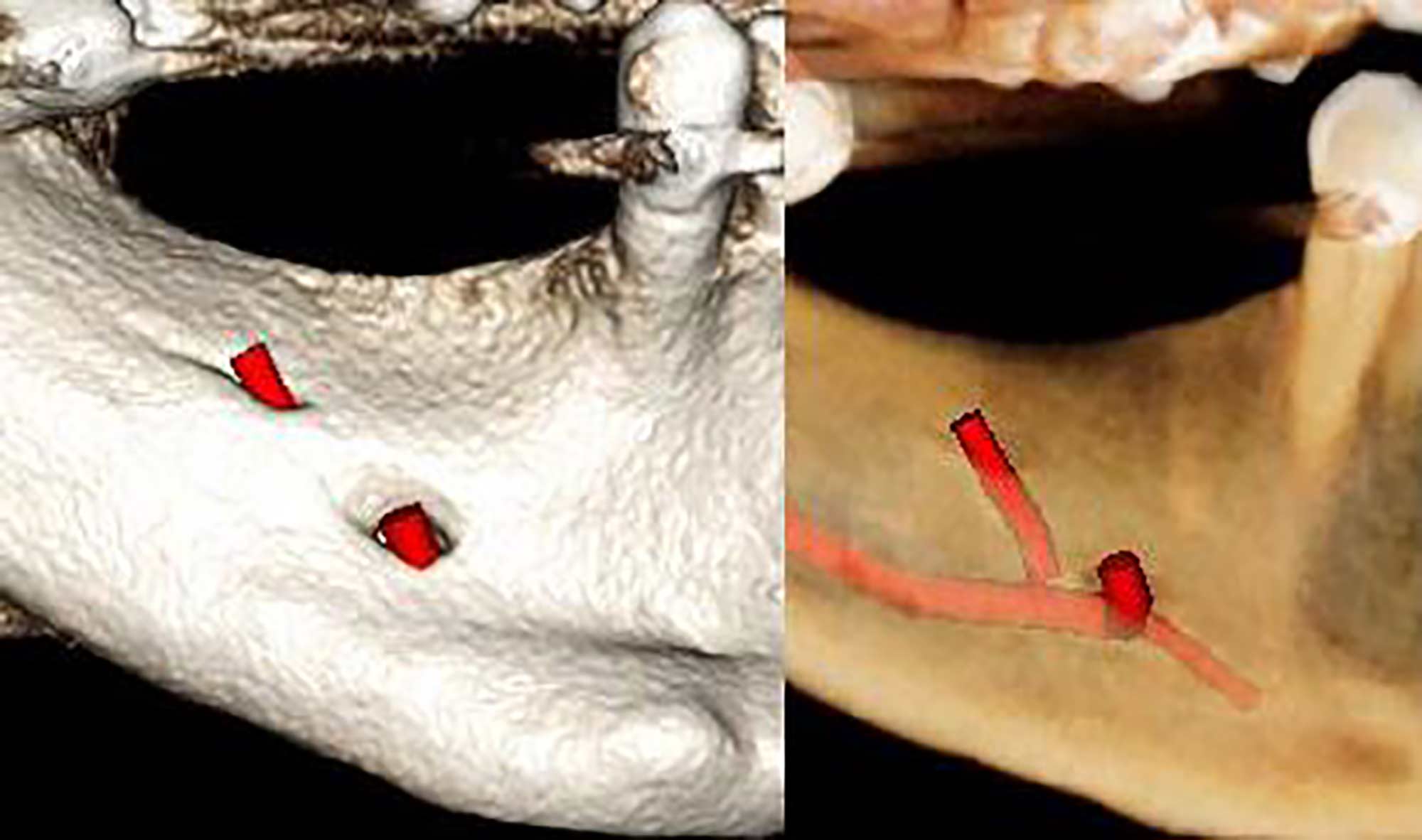
The findings (Figures 1 and 2) showed the right mandible exhibits two foramina, which is an uncommon variation in normal anatomic form.
All CT Dent cases are available to view and manipulate online on the company’s cloud viewer software. For further cases, visit https://ct-dent.co.uk/account/cases/.

