Dentistry
Dentistry’s guide to digital dental X-rays
Last updated: 26th May 2026
Contents
Digital dental X-rays are increasingly being embraced by dental professionals as they move away from traditional X-rays. Digital dental radiography may seem expensive and difficult to adapt to, but it is possible to offset the initial costs over time. Digital X-rays can save money on the cost of developing film, while providing faster and more effective imaging.
Traditional X-rays were discovered in 1895, and it was only in 1987 that the first digital dental X-ray was made available. By 2024, the UK market was worth an estimated $583.63 and is expected to reach £837.49m by 2029.
In this guide, we will explore the types of digital X-ray machines, how they work and some of the leading manufacturers.
What is a digital dental X-ray?
Like traditional X-rays, digital dental X-rays use electromagnetic waves, which pass through human tissue and are detected by a receptor behind it.
But rather than using photographic film to record the image, digital X-rays use digital sensors that feed the image into a computer. The image is then immediately available for review, without need for developing.
What are the benefits of digital dental radiography?
There are numerous reasons for moving towards digital dental X-rays for you and your patients.
- Higher quality images: image resolution is typically higher for digital dental X-rays, thanks to the sensors they use. This allows images to be enlarged and otherwise manipulated to a far greater extent
- Fewer waste products: chemicals used in traditional X-ray process can cause environmental damage, as can discarded film. Digital X-ray technology uses neither, making it far more sustainable
- Less radiation: digital dental X-rays can use just 10% of the radiation of traditional X-rays. This makes them far less harmful for patients and practitioners
- Image manipulation: dental digital radiography allows dentists to alter images’ brightness, contrast and other variables. This can make it easier to see elements of the image
- Storage: traditional dental X-rays demand a large amount of space. Physical X-ray images need to be kept somewhere. However, digital radiography images can be saved to a hard drive, network or on the cloud. They can also be reproduced and transmitted to other medical professionals with ease
- Costs: digital dental X-ray machines can cost tens of thousands of pounds. It might therefore seem counterintuitive to suggest that they are cheaper than traditional X-rays. However, there are long-term savings to be made, such as the costs involved with developing film. The speed and efficiency of modern digital X-ray machines also mean faster treatments, allowing you to see more patients.
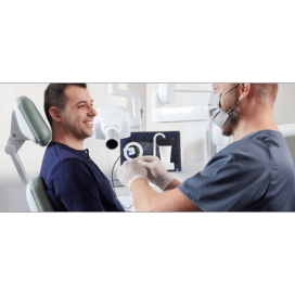
What types of digital dental X-rays are there?
Digital dental X-rays fall into two main categories, intraoral and extraoral:
Intraoral X-rays
Intraoral X-rays (IOX) are taken inside the mouth. There are three main types.
- Bitewing X-rays: bitewing dental X-rays are used to see the upper and lower teeth in an area of the mouth. Bitewing X-rays can be used to see decay between teeth, determine bone density and fit dental crowns and restorations. They can also discover mouth cancers, orthodontic problems and infections. For these, the patient bites down on a sensor, holding it in place while the X-ray is taken. The image shows teeth from the crown to the base. They do not usually show the root
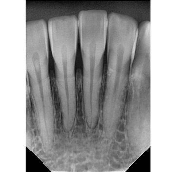
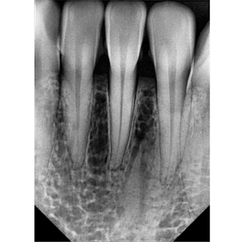
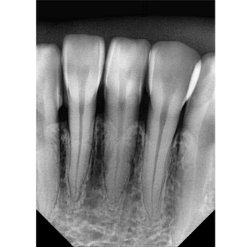
- Periapical X-rays: periapical X-rays are used to see a tooth’s root structure and surrounding bone structure. They can determine bone loss, decay and gum disease. Abnormalities to teeth and their surrounding bone can also be observed. This also involves the patient biting down on a sensor while the X-ray is taken
- Occlusal X-rays: occlusal X-rays are used to image the floor of the mouth or the roof/palate. These provide images of tooth and bone structure for the entire mouth. Patients are required to hold the sensor or film between upper and lower teeth for the X-ray to be taken.
Extraoral X-rays
Extraoral X-rays are taken from outside the mouth. They include:
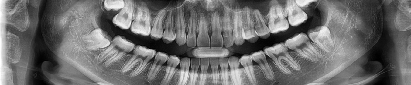
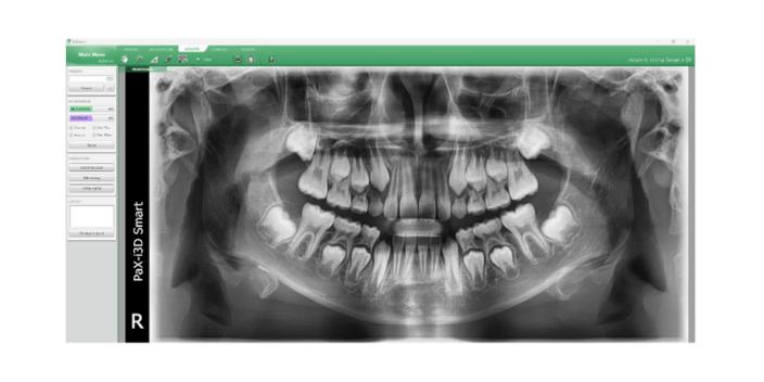
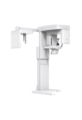
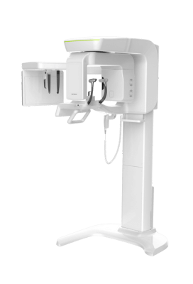
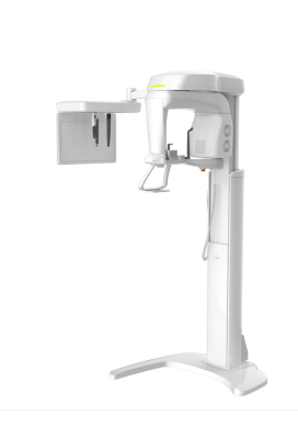
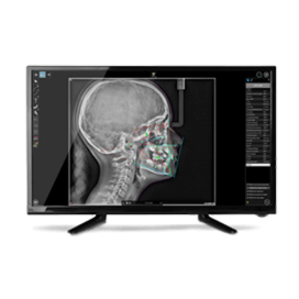
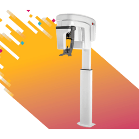
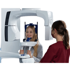
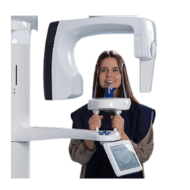
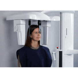
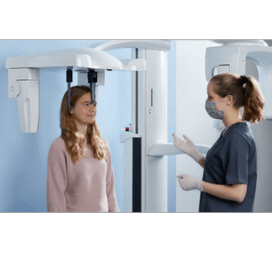
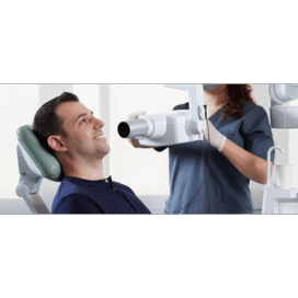
- Panoramic/OPG X-rays: panoramic X-rays, also known as panorex or orthopantomogram (OPG) X-rays, show the whole mouth in a single image. This includes all teeth in the upper and lower jaws and surrounding bone and tissue. Uses of panoramic X-rays include the diagnosis of jaw problems, impacts and impacted wisdom teeth. They are also used to detect cysts, jaw tumours and oral cancer. The image is taken using a rotating arm that moves around a patient’s head in a semi-circle
- Lateral cephalometric projections: lateral cephalometric projections take images of the entire head. They are mainly used for orthodontic diagnosis and to plan treatments
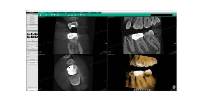
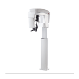
- Cone beam computerised tomography: cone beam computerised tomography (CBCT) is used to image the internal structures of the head in three dimensions. It can be used to identify problems in facial bones, fractures and tumours. It can also define the exact structure of the jawbone during the dental implant process. During CBCT imagining, the patient sits or stands while the scanner rotates around the head.
What to look for in a digital dental X-ray machine
It is important to choose the right digital X-ray machine for you. Here are some factors to consider:
Machine quality
A high-quality X-ray machine means fewer breakdowns and less downtime. This means it will last longer, avoiding the cost of a replacement. Quality is also important in terms of ensuring your machine is operating safely.
Image quality
There remains a significant difference in image quality between 3D and even 2D X-ray images.
It is essential to determine whether the superior image quality is a result of advanced software tools or the inherent quality of the images themselves. The voxel size plays a crucial role in producing high-resolution 3D images. It directly affects the detail and accuracy of the output.
When investing in X-ray technology, prioritise systems that offer low-dose X-rays without compromising on image quality. Achieving this balance is vital for accurate diagnostics and enhanced patient care.
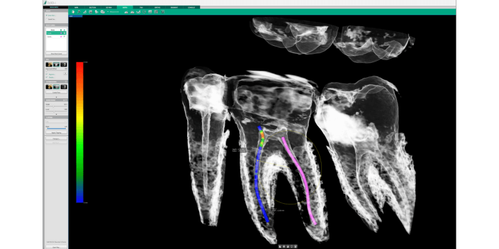
In particular, voxel size is a game-changer in endodontic treatment.
High-resolution 3D images, enabled by optimal voxel size, allow for precise visualisation of root canal anatomy. This clarity is crucial for effective diagnosis and treatment planning, ultimately enhancing the success rate of procedures.
Costs
The costs associated with buying a new digital X-ray machine are more than just its price tag. For a start, you will need to install it, which may also involve changes to your surgery’s design. X-ray rooms will need to be made compliant with UK Health Security Agency and Radiation Protection Adviser guidelines. You may also need to pay for maintenance, repairs and parts in the future.
Investing in digital X-ray machines differs significantly from other types of equipment, especially when it comes to patient billing.
This investment is often viewed through the lens of ‘return on investment’ (ROI). Unlike traditional machines, digital X-ray systems allow for enhanced efficiency and quicker diagnosis. This ultimately leads to improved patient care.
Moreover, the ability to charge patients directly for these services can enhance revenue streams. Thus, understanding the ROI associated with digital X-ray machines is crucial for making informed purchasing decisions.
This strategic investment not only benefits the practice financially but also elevates the quality of service provided to patients.
Warranty
The warranty on X-ray machines is important, as it directly impacts costs.
Without a comprehensive warranty, you could end up spending thousands of pounds on repairs.
Therefore, it’s essential to clarify the type of warranty offered – whether it is a full parts warranty or limited to main components. Understanding these details will help you make a more informed decision and protect your investment in the long run. A robust warranty safeguards against unforeseen costs.
Customer support
Reliable customer support is invaluable for promptly addressing any issues that may arise. Prioritising ease of use of both the machine and software will ultimately improve your practice’s efficiency and patient care.
Software usability and training
It is crucial to ensure that both the machine and the imaging software are user-friendly.
While capturing an X-ray may seem straightforward, you might encounter challenges with software that is not intuitive. This can lead to frustration and inefficiencies in your workflow.
Therefore, seek out a provider that offers comprehensive user training and robust local customer support. Having access to excellent training resources can significantly enhance your team’s proficiency and confidence in using the equipment.
Safety
Different models of digital dental X-ray machine emit different levels of radiation. The level of radiation required to capture a quality image will vary between machines. Keeping radiation exposure to a minimum is vital to the health and safety of your patients and staff. Modern digital X-ray machines often require lower doses of radiation.
Given that all X-rays involve radiation, choosing low-dose X-ray technology is important, especially when treating children.
Children are more sensitive to radiation exposure, so utilising low-dose options can significantly reduce potential risks. This approach not only ensures their safety but also provides peace of mind for parents and caregivers. When selecting X-ray equipment, consider the technology’s ability to minimise radiation while still delivering high-quality imaging.
Digital dental X-rays buying guide
Vatech

Vatech is the only CBCT specialist in the market that designs, produces and manufactures core components in-house. It does this at its state-of-the-art facility in South Korea. This allows Vatech to offer a standard five-year full parts warranty, which can be extended to 10 years.
Vatech stands out as the only company in the market that reinvests up to 25% of its sales into research and development (R&D).
This commitment to innovation has led to a remarkable history of groundbreaking achievements. For instance, Vatech introduced the world’s first three-in-one CBCT and auto-switching CBCT systems.
Furthermore, more than a decade ago, Vatech was the first to develop Green CBCT technology, ushering in the era of low-dose dental X-rays. It pioneered the development of Green CBCT technology, dual-scanning CBCT and flexible sensors. Additionally, Vatech has embraced cutting-edge advancements, such as carbon nano technology adaptations in intraoral X-rays and the creation of a 49.5-micron voxel endo CBCT.
This relentless pursuit of excellence not only demonstrates Vatech’s dedication to improving imaging technology. It also ensures that practitioners have access to the most advanced tools for providing high-quality patient care.
Vatech offers the most precise imaging in the industry, with a voxel size of 49.5 microns.
Vatech’s extraoral X-rays
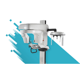
CBCT
Vatech Green X and Smart X offer the best image quality on the market with the smallest voxel size. They offer advanced imaging capabilities for a wide range of dental procedures.
Green X

The Green X has five distinct innovative features:
1: The Green X Endo mode, with the world’s smallest voxel size of 49.5 microns, allows clinicians to obtain more detailed information. This increases their chances of solving clinical problems.
2: Its Ez3D-I Endo software supports the whole process of surgery. Itsc has specialised functions for diagnosis, implant planning, simulation and consultation, especially endodontic plan with root canal tracing.
3: Vatech’s green technology reduces the radiation level of the Green X without reducing image quality with Green Scan Time.
4: Advanced metal artefact reduction technology enhances image clarity. It improves diagnostic accuracy by minimising the distortions, or artefacts, caused by metal objects. These include dental implants, crowns, braces and surgical hardware.
5: Magic pan technology produces superior panoramic images by eliminating distortions and blurring caused by improper patient positioning.
Smart X
Smart X is the latest addition to Vatech’s premium X series. It is thoughtfully designed to incorporate the innovative features of the Green X while offering enhanced cost efficiency.
The world’s smallest voxel size
Smart X also has Endo mode, with the world’s smallest 49.5 μm voxel size. This enables clinicians to access more comprehensive information, enhancing their ability to address clinical challenges effectively.
Smart focus mode
Smart focus mode allows for ultra-high-definition images at a 4x4cm size within a 12x9cm FOV using endo mode. It is powered by Vatech’s world-leading X-ray generator and sensor technology and combined with AI-based software.
Sleek and modern design
Smart X stands out for its sleek, ergonomic and modern design. Minimalist and sophisticated, it seamlessly integrates into clinical environments while maximising space efficiency.
Smart Plus
Smart Plus has a three-in-one digital X-ray imaging system that incorporates pano, ceph (optional) and CBCT (with auto pano). The Smart Plus provides high-quality images with lower radiation.
One scan, two images
One scan with the Smart Plus gives you a CT image and an auto pano image. This means, patients who require both images do not need to undergo two X-ray scans.
Anatomical FOV 12X9
The innovative FOV of the Smart Plus provides an arch-shaped volume. This shows a wider view of dentition compared to other devices of the same FOV.
Compressed sensing technology
Vatech dramatically improves its image quality with much less artefact and noise via its compressed sensing technology (CST). This iterates its reconstruction process 10 times more than the normal amount to depict the object’s true representation.
OPG
Pax-I Plus
The Pax-I Plus provides the most precise and high-quality panoramic images by combining image processing.
Vatech’s intraoral X-rays
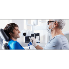
Currently, the X-rays drawing the most attention from dentists are handheld and wall mounted. This is because they allow for immediate imaging in the surgery room and are priced more affordably compared to CBCT.
The most trusted handheld X-ray available on the market today is Vatech’s Ezray Air Portable.
Ezray Air Portable
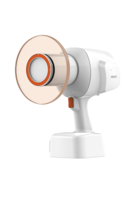
The Ezray Air Portable is the world’s first X-ray device utilising carbon nano technology (CNT).
To learn more about CNT, please check out this video.
Lightweight
At 1.8kg, the Ezray Air Portable is a lightweight portable X-ray device designed for easy handling and stable positioning. It delivers optimal image quality for your intraoral X-ray images. The ease of device positioning produces clear images without any motion artefact.
No warmup, faster cooling
Unlike conventional X-rays, Ezray Air Portable has no start-up delay as the source warms up. Its carbon nano technology allows for quicker exposure after you turn on the device.
Ezray Air Portable has an almost 75% reduction in cooldown time between shots when compared to leading competitor devices.
Double shield: internal shielding and external backscatter shielding
The internal radiation shielding is perfectly designed to protect the operator from radiation leakage. Exposure to radiation results from the beam interacting with the surface of the patient. This causes radiation to bounce off as radiation scatters in different directions. The backscatter shield significantly reduces the amount of radiation being reflected.
Ezray Air has been approved by the European Commission (EC), the International Organisation for Standardisation (ISO) and the Food and Drug Administration (FDA), ensuring its safety and reliability.
Ezray Air Wall
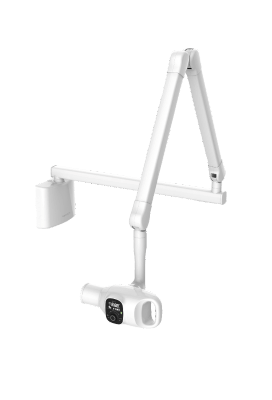
Lightweight
The Ezray Air Wall, a wall-mounted unit featuring carbon nano technology, is designed to be lightweight. Its compact tube head offers a stable and user-friendly X-ray source that enhances image clarity and streamlines workflow.
This intraoral X-ray is fully equipped for rigorous daily use. It is crafted to be simple for dentists to operate and easy to position accurately while requiring minimal space.
Ergonomic design for safety and efficiency
- Round shape design: the Ezray Air adds a modern and chic touch to your clinic
- Strong and stable arm: the strong and stable X-ray unit arm allows accurate positioning of a 2.4kg lightweight tube head, aiding distortion-free images
- Ergonomic one-handed grip: a hand grip provides an easy method of positioning the Ezray Air quickly and accurately.
Safety and reliability
Ezray Air Wall has been approved by the EC, ISO and FDA, ensuring its safety and reliability.
Intraoral sensors
IOX should be used in conjunction with either an intraoral sensor or a phosphor plate.
The Ezsensor Classic and Ezsensor HD are Vatech’s flagship intraoral sensors. They are available in sizes suitable for children (1.0 size), adults and a 1.5 size that can be used by both adults and children. Its rounded design ensures comfortable use for patients.
Photostimulable phosphors
VSP
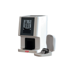
Vatech VSP is one of the smallest and lightest photostimulable phosphors (PSP) scanners. It weighs only 3.3kg and has dimensions of 233x141x191.5mm.
VSP output image quality is always in super resolution (20lp/mm) or high resolution (10lp/mm).
The world’s first PSP scanner was created using multi-pixel photon counters (MPPC). What sets MPPC apart from the existing photo multiplier tube (PMT) is immunity to magnetic fields and high resistance to mechanical shock.
Also, the MPPC is lower than 10 times voltage operation. And more than that, 10 times high gain can be obtained when compared to PMT. And it is a small size. MPPCs can be driven at a low voltage, are small in size and are inexpensive.
Store up to 500 high-resolution images
The Vatech VSP has a built-in memory, so you can store up to 500 scan images without a PC connection. It is possible to verify that imaging is done normally through the LCD preview screen even when scanning without PC.
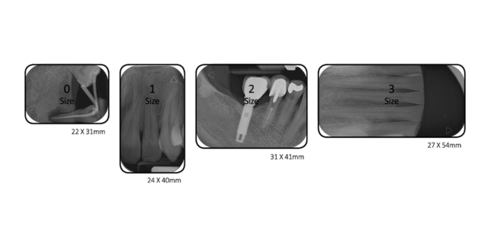
Four sizes
Flexible imaging plates come in four sizes. Cableless, thin and soft imaging plates optimally stick to various oral shapes to provide high quality images while maintaining maximum comfort for the patient.
Image plates are highly durable and reduce the risk of damage. Ultra-thin imaging plates can be reused repeatedly while maintaining great image quality.
Vatech offers…
Reliable 10-year warranty
Vatech offers a five-year full parts warranty, extendable by another five years, for a total of ten years. It is the only X-ray manufacturer that designs and manufactures all core components, at its headquarters in South Korea.
It can swiftly and efficiently provide any parts needed for repairs. Our products are built to last, which is why we confidently offer warranty coverage for up to 10 years.
Customer service and training
Vatech’s customer service and comprehensive user training are widely recognised as second to none. Vatech UK is proud to have three in-house X-ray experts, supported by our European headquarters in the Czech Republic.
Vatech also offers a specialised CBCT interpretation course for dentists.
Free CBCT consultation
Vatech offers a wide range of CBCT products. It also provides free consultations to help customers choose the CBCT product most compatible with their speciality and treatment programmes. It also provides detailed guidance throughout the purchase and installation process.
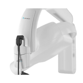
Acteon

With panoramic imaging systems, CBCT and intra-oral imaging, Acteon offers a comprehensive product portfolio. Acteon Imaging, manufactured by Sopro and de Götzen, promotes digital dental imaging systems and dental radiology.
Intraoral X-rays
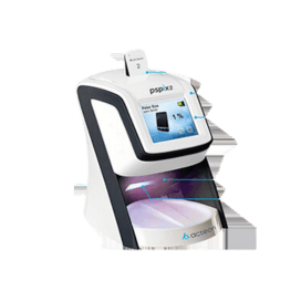
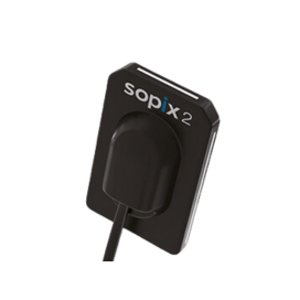
Pspix 2 has a fully automated process. Drop the plate into the slit and the Pspix2 will automatically detect the size, scan the plate, optimise the image and send it instantly to your imaging software. It has a resolution of 20 lp/mm.
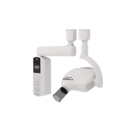
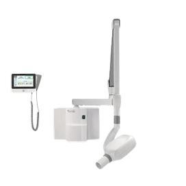
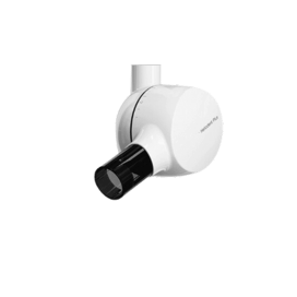
The X-Mind unity intraoral X-ray reduces radiation exposure. It has an elegant style and cutting-edge patented automatic exposure control (ACE) technology. It can be integrated with the Sopix plug-in sensor.
ACE technology analyses in real-time the amount of X-rays accumulated by the Sopix digital X-ray sensors. It freezes image acquisition when the sensor receives the radiation dose to produce an optimal image. It has a real resolution of >18 lp/mm.
Combined with the X-Mind Unity intraoral X-ray generator, the Sopix plug-in with ACE technology limits the emission of X-rays during acquisition to the necessary amount for the patient’s morphology. It has a real resolution of >18 lp/mm.
Cephalometry
The ergonomics of X-Mind Prime Cep’s teleradiography head support is designed to optimise patients’ comfort and stability. It aims to reduce the risk of retakes because of motion artefacts. Its patented blade collimator provides a wide range of image formats to fit almost any clinical need.
Panoramics/OPG
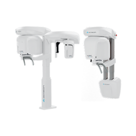
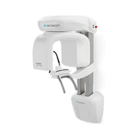
X-Mind Prime is a new-generation digital extra-oral dental unit. It has a stylish and smooth design underlying a cutting-edge technology. It is also available with a ceph arm. Its voxel size is 87.5 microns.
CBCT
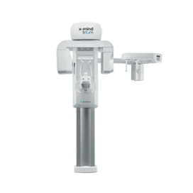
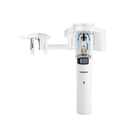
X-Mind Trium True Low Dose offers a radiation dose reduction of up to 50%, without a drop in quality. The new specific acquisition mode for smaller patients, particularly children, reduces radiation exposure.
X-Mind Prime 3D is a new generation CBCT all-in-one dental unit. It offers a stylish and smooth design and a streamlined acquisition workflow.
Belmont

Belmont is a leading manufacturer of treatment centres, operating lights and X-ray machines that facilitate better treatment experiences for dentists and patients.
Intraoral
Belmont Touch features touchscreen controls. It has a handheld exposure switch to control radiation doses. The device can be kept in sleep mode when inactive. It has three preset settings, for adults, adolescents and children. It has a range of models to suit different practices.
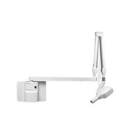
Belmont’s 097 Belray II intraoral X-ray is safe and simple to operate. It features adult, adolescent and child presets, which can be manually overridden for more challenging exposures. It can also easily switch between film and digital imaging systems.
Carestream

Carestream Dental aims to simplify cutting-edge technology in order to transform the dentistry profession. Headquartered in Atlanta, Georgia, its areas of focus include supplying cloud-based technology solutions for dental practices.
Extraoral
CBCT

The CS 8200 3D Access promises crystal-clear 3D images that are easy to obtain. It is state-of-the-art while being intuitive to new users. CS Upstream premium support is included.
The CS 8200 3D Neo Edition has the broadest range of fields of view in its category. It offers full control of the imaging area using its Scout View function. The CS 8200 3D Neo Edition CBCT system is the most advanced in the CS 8200 3D family.
The CS 9600 is a high-precision five-in-one CBCT scanner. It uses artificial intelligence positioning to ensure consistent results. It also has a broad range of volume sizes, with up to 14 fields of view. It now also has a Scan Ceph module, among other innovations. The CS 9600 CBCT scanner is ideal for precision.
Cephalometric
Carestream Dental’s imaging systems are designed for accuracy and speed. They offer ultra-fast scanning technology and automatic tracing software.
Panoramic
The CS 8100 Evo Edition utilises Tomosharp technology and powerful image processing. It is designed to be intuitive to use and easy to install, using its ‘plug-and-pan’ functions. The CS 8100 Evo Edition delivers high-quality, clear images, with optimal sharpness and contrast. It is also ultra compact, meaning it can fit into small spaces.
Intraoral sensors
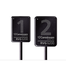
The RVG 6200 offers a simplified workflow for image processing tools. The CS Adapt module lets users choose from 40 pre-set image enhancement filters or define up to four their own favourite settings. It also offers three anatomical image enhancement modes. These can be applied to endodontic, periodontic and dentin-enamel junction images. It has a sharpness filter, and a dynamic slider bar shows contrast changes in real time.
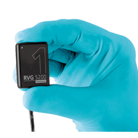
The RVG 5200 is suited to those taking their first step into digital dental radiography. The image quality of the RVG 5200 sensor is the same or higher than other intraoral sensors in its class. It features three anatomical image enhancement modes, for endodontic, periodontic and dentin-enamel junction images. It has a sharpness filter, and a dynamic slider bar shows contrast changes in real time.
Intraoral generators for dental X-rays
The CS 2200 features a very high-frequency (DC) X-ray generator. This results in high image quality and reproducibility. The CS 2200 is also designed to improve safety conditions. Its timer determines appropriate exposure times based on anatomical region, patient bodyweight and detector type.
The CS 2100 is an affordable high-frequency technology generator. It optimises contrast and image definition. It works with both film and digital radiography systems. The 60kV generator has a 0.7mm focal spot for sharp, contrasted images. The exposure time can be optimised as needed.
Dentsply Sirona
US-headquartered Dentsply Sirona invests more than $125 million a year in advancing its dentistry technology and innovation. It works with practices, clinics and dental laboratories around the world, and has over 4,000 staff providing customer service.
Extraoral
Axeos is Dentsply Sirona’s most advanced CBCT X-ray system. It has a wide range of volume sizes up to Ø 17x13cm. Axeos can be used for myriad diagnostic and treatment planning needs.
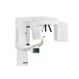
The Orthophos S is designed for usability for dentists looking to enhance patient care and grow their practices. It boasts excellent image quality and advanced features. It can be used to diagnose and create treatment plans by capturing everything from a single tooth to the airway.
Orthophos E is a solid 2D X-ray unit designed to provide a smooth entrance into the world of digital extraoral imaging. It has an optional left ceph arm.
Intraoral

Xios XG Supreme is an intraoral X-ray sensor system for digital X-rays in HD quality.
Heliodent Plus is a reliable and flexible intraoral X-ray generator, which can be used for optimised diagnostics.
Kavo

Kavo is one of the world’s most successful dental manufacturers. It has been a driving force in innovations for more than 110 years.
Extraoral
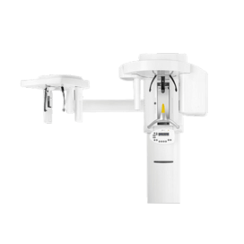
The Kavo Proxam 3DQ X-ray unit can be used for a variety of 3D diagnostic options. It is one of Kavo’s premium products and brings together the qualities of Kavo Proxam 3D and 2D imaging. Thanks to its preconfigured programmes, it offers a wide range of diagnostic capabilities and ENT applications.
Kavo Proxam 3D brings together maxillofacial 3D imaging with 2D imaging options. It offers programmes to suit children and bitewing and cephalometric X-rays. It uses low levels of radiation to create detailed dental volume tomography images.
Kavo Proxam 2D is designed to bring together a host of diagnostic needs in the dental surgery. These include panoramic, extraoral bitewing, temporomandibular joint and sinus images. It offers easy to use versatile extraoral X-ray imaging at high quality.
Ceph imaging
Ceph imaging with Kavo Proxam is specifically designed for orthodontics. It supports a variety of image formats and offers image sizes of up to 30x27cm.
It can be used as an additional feature for all extraoral Kavo Proxam imaging units. It uses low levels of radiation due to its narrow X-ray beam, which scans patients’ heads horizontally.
Intraoral
The Kavo Proxam IX creates intraoral X-rays at the highest level. It offers intuitive operation, precise positioning and a simple imaging process. It uses a small focal point and allows variable exposure parameters to create high quality 2D images.
Kavo Proxam IS sensors produce high quality images in a short space of time. They produce high-definition images at all doses of radiation. They are also durable and comfortable to use.
Kavo Proxam IP is an imaging plate scanner for use anywhere in the practice. It allows fast, easy and uncomplicated workflows. The Kavo Proxam IP’s featured include its ability to image large scan areas with full exposure and automatic error avoidance. It is also quiet and compact.
The Kavo Proxam IOS intraoral scanner creates high-speed and high-precision scans of excellent quality. It is fully integrated with Planmeca Romexis software.
Conclusion
Digital X-rays offer a host of benefits when compared to traditional X-rays. Quality, detail, sustainability and the ability to manipulate and share images can hugely improve your offering.
This guide has shown the range of functions available in modern digital dental X-rays and some of the devices on the market.
Think about your needs, budget, available space and other factors when making your choice. The right decision could transform your practice.
Stay updated with relevant information about this webinar