Dr Matthew R Miller presents an introduction to his white paper focusing on the management of complex root canal anatomy, comparing Kerr’s Zenflex file system with two other popular systems.
Dr Matthew R Miller graduated from New York University (NYU) college of dentistry with high distinction.
While at NYU, he was selected into the honours aesthetics programme in the Rosenthal Institute of Cosmetic Dentistry.
As a result of his efforts, he won the American Academy of Aesthetic Dentistry Award.
Matthew’s scope of practice includes full mouth rehabilitation, endodontic therapy, Invisalign, oral surgery, bone grafting, sinus augmentation, and implant therapy.
He is also a key opinion leader for Kerr and is a member of their endodontic and restorative advisory board.
Additionally, he is a clinical consultant and evaluator for the Dental Advisor. He maintains a private group practice in North Carolina, USA.
Core principles
Non-surgical endodontic therapy can be achieved by various filing and obturation methods.
However, there are certain core principles to which one must adhere.
The canal system must be debrided of bacteria and contaminants. Also, it must be disinfected, and the root apex must be appropriately sealed along with the coronal aspect of the tooth.
In order to perform this therapy successfully, the clinician must have a proper foundation in each of these principles and must comprehend tooth morphology and associated varying complexities.
Weine, Vertucci and Ahmed (1969), among others, established classification systems for root canal morphology that denote the commonly occurring canal configurations (Table 1).
But the challenge still remains in how to best manage and treat these complex anatomical variances.
Several factors, such as the number of roots, the number of canals, canal classification, presence of stenosis and or calcifications, sharp curvatures, lateral canals, and atypical apices, can affect the success rate of non-surgical endodontic therapy.
And while even though the use of proven clinical techniques and sealers can still result in failed endodontic therapy, advancements in techniques and materials are helping to mitigate this incidence of failure.
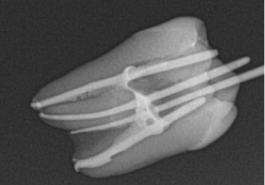
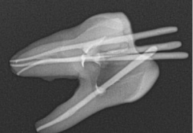
CBCT imaging has also played a major role in the advancement of endodontics by allowing clinicians to diagnose cases more effectively, as well as identify and recognise tooth and canal morphology and anatomy.
For the past 30 years, a combination of hand files and nickel titanium rotary files have been used to instrument and debride the canals to then allow for delivery of disinfecting irrigants into the canal system and subsequent obturation.
The right technique
A widely accepted technique is to pre-enlarge the coronal and middle thirds of the root to then perform scouting and final instrumentation of the apical third.
This technique allows for adequate debris removal, introduction of irrigants, with less risk of the file binding in multiple points. This can lead to separation and or potential for intra-canal fractures.
And while the metallurgy of these NiTi (nickel titanium) rotary files allow them to be stronger and more flexible than their stainless-steel counterparts, they can still be susceptible to breakage, even without prior use or signs of deformation.
Utilisation of nickel titanium files with this technique is still considered to be advantageous over stainless steel. However, some of the risks and disadvantages to using them in this method include:
- Over instrumentation and removal of dentine, leading to thinning of the root canal walls and potential for subsequent fracture
- Straightening and transportation of canal and apex
- Ledging within the canal
- Extrusion of debris
- Apical blockage
- File separation.
Current concepts
Current concepts of endodontic therapy place an emphasis on glide path, conservative dentine removal and flexibility in their file designs in an attempt to preserve as much as the natural tooth anatomy as possible.
Glide path can reduce these aforementioned risks by facilitating canal shaping during the instrumentation process while following the existing path of the canal.
Canal anatomy and curvatures vary greatly, and curvatures greater than 30 degrees are considered significant and can pose significant challenges and risks. These can include file separation, canal straightening and transportation, ledging, and stripping and perforation.
Because of this, glide path management can be a crucial step in the outcome of endodontic therapy.
Many manufacturers have developed thermally treated file systems to perform endodontic therapy conservatively.
This is done by designing efficiently cutting, safe-ended, flexible files with an offset centre of rotation, controlled memory, and a maximum flute diameter.
This allows the files to follow the natural anatomy and curvature of the canal.
At the same time, these improvements reduce the risk of file separation due to cyclic fatigue and torsional stress.
The goal of these modifications is to improve the overall success of endodontic therapy, provide efficient cutting, preserve the natural shape of the canal, and resist fracture of the file even in the most demanding of circumstances, such as narrow and curved root canals.
Sufficiently instrumented canals allow irrigants to be introduced and activated for three dimensional disinfection and subsequent obturation.
Optimal, minimally invasive endodontic therapy
Following a comparative review of three file systems, each having different characteristics and approaches for treating complex anatomy, the author was highly impressed by Kerr’s Zenflex file system’s ability to treat all cases endodontically, from simple to complex, using the clinician’s preferred technique.
The author especially loved how it preserves the integrity of the tooth structure and natural anatomy with its controlled memory and one millimetre maximum flute diameter.
The proprietary variable heat treatment based on mass makes these files incredibly strong and flexible. It also provides exceptional cutting efficiency, and significantly reduces the risk for separation.
With Kerr Zenflex files, clinicians can provide optimal, minimally invasive endodontic therapy.
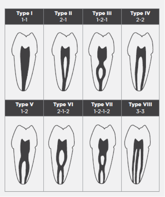
Canal classification table
- Type I: a single canal extends from the pulp chamber to the apex
- Type II: two separate canals leave the pulp chamber and join short of the apex to form one canal
- Type III: one canal leaves the pulp chamber, divides into two within the root, and then merges to exit as one canal
- Type IV: two separate and distinct canals extend from the pulp chamber to the apex
- Type V: one canal leaves the pulp chamber and divides short of the apex into two separate and distinct canals with separate apical foramina
- Type VI: two separate canals leave the pulp chamber, merge in the body of the root, and re-divide short of the apex to exit as two distinct canals
- Type VII: one canal leaves the pulp chamber, divides and then rejoins within the body of the root, and finally redivides into two distinct canals short of the apex
- Type VIII: three separate and distinct canals extend from the pulp chamber to the apex.
For further information about Kerr Zenflex files, please visit store.kerrdental.com/en-uk.

